Banzato Tommaso, Bonsembiante Federico, Aresu Luca, Zotti Alessandro
Department of Animal Medicine, Production and Health, Clinical Section, Radiology Unit, University of Padua, Viale dell'Università 16, Legnaro, 35020, Padua, Italy.
Department of Comparative Biomedicine and Food Science, University of Padua, Viale dell'Università 16, Legnaro, 35020, Padua, Italy.
BMC Vet Res. 2017 Jan 17;13(1):24. doi: 10.1186/s12917-016-0941-z.
Renal cortical echogenicity is routinely evaluated during ultrasonographic investigation of the kidneys. Both in dog and cat previous ex-vivo studies have revealed a poor correlation between renal echogenicity and corresponding lesions. The aim of this study was to establish the in-vivo relationship between renal cortical echogenicity and renal histopathology.
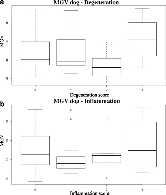
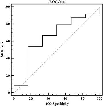
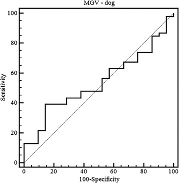
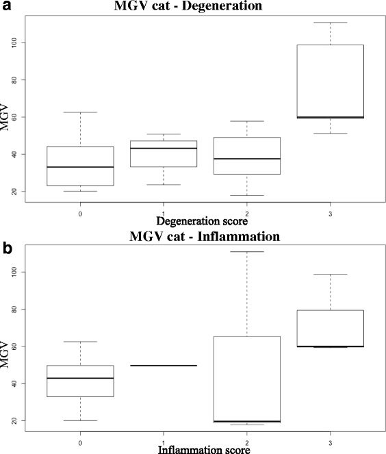
Thirty-eight dogs and fifteen cats euthanized for critical medical conditions were included in the study. Ultrasonographic images of both kidneys were acquired ante mortem at standardized ultrasonographic settings. The echogenicity was quantified by means of Mean Gray Value (MGV) of the renal cortex measured with ImageJ. A complete histopathological examination of both kidneys was performed. Five kidneys were excluded because histopathology revealed neoplastic lesions. Only samples affected by tubular atrophy showed statistically different values in dog, and histopathology explained 13% of the total variance. MGV was not correlated neither to the degeneration nor to the inflammation scores. However, significant differences were identified between mildly and severely degenerated samples. Overall, the classification efficiency of MGV to detect renal lesions was poor with a sensitivity of 39% and a specificity of 86%. In cats, samples affected by both tubular vacuolar degeneration and interstitial nephritis were statistically different and histopathology explained 44% of the total variance. A linear correlation was evident between degeneration and MGV, whereas no correlation with inflammation was found. Statistically significant differences were evident only between normal and severely degenerated samples with a sensitivity of 54.17% and a specificity of 83.3% and MGV resulted scarce to discriminate renal lesions in this species.
Renal cortical echogenicity shows low relevance in detecting chronic renal disease in dog whereas it results worth to identify severe renal damage in cat.
在肾脏超声检查中,通常会评估肾皮质回声。此前针对犬猫的体外研究均显示,肾回声与相应病变之间的相关性较差。本研究旨在建立肾皮质回声与肾脏组织病理学之间的体内关系。
本研究纳入了38只因严重疾病而实施安乐死的犬和15只猫。在标准化超声检查条件下,于安乐死前获取双侧肾脏的超声图像。通过ImageJ测量肾皮质的平均灰度值(MGV)来量化回声。对双侧肾脏进行了完整的组织病理学检查。5个肾脏因组织病理学显示存在肿瘤性病变而被排除。仅受肾小管萎缩影响的样本在犬中显示出统计学差异,且组织病理学解释了总方差的13%。MGV与变性评分和炎症评分均无相关性。然而,在轻度和重度变性样本之间发现了显著差异。总体而言,MGV检测肾脏病变的分类效率较差,敏感性为39%,特异性为86%。在猫中,受肾小管空泡变性和间质性肾炎影响的样本存在统计学差异,且组织病理学解释了总方差的44%。变性与MGV之间存在明显的线性相关性,而与炎症无相关性。仅在正常样本和重度变性样本之间存在统计学显著差异,敏感性为54.17%,特异性为83.3%,MGV在该物种中难以鉴别肾脏病变。
肾皮质回声在检测犬慢性肾病方面相关性较低,而在识别猫的严重肾损伤方面具有一定价值。