• 文献检索
  • 文档翻译
  • 深度研究
  • 学术资讯
  • Suppr Zotero 插件Zotero 插件
  • 邀请有礼
  • 套餐&价格
  • 历史记录
应用&插件
Suppr Zotero 插件Zotero 插件浏览器插件Mac 客户端Windows 客户端微信小程序
定价
高级版会员购买积分包购买API积分包
服务
文献检索文档翻译深度研究API 文档MCP 服务
关于我们
关于 Suppr公司介绍联系我们用户协议隐私条款
关注我们

Suppr 超能文献

核心技术专利:CN118964589B侵权必究
粤ICP备2023148730 号-1Suppr @ 2026

文献检索

告别复杂PubMed语法,用中文像聊天一样搜索,搜遍4000万医学文献。AI智能推荐,让科研检索更轻松。

立即免费搜索

文件翻译

保留排版,准确专业,支持PDF/Word/PPT等文件格式,支持 12+语言互译。

免费翻译文档

深度研究

AI帮你快速写综述,25分钟生成高质量综述,智能提取关键信息,辅助科研写作。

立即免费体验

剪切波弹性成像和超声造影在甲状腺恶性结节诊断中的应用

Shear wave elastography and contrast-enhanced ultrasonography in the diagnosis of thyroid malignant nodules.

作者信息

Chen Mei, Zhang Ke-Qin, Xu You-Feng, Zhang Sheng-Min, Cao Yong, Sun Wei-Qun

机构信息

Department of Ultrasonography, Ningbo First Hospital, School of Medicine, Zhejiang University, Ningbo, Zhejiang 315000, P.R. China.

Department of Endocrinology, The Affiliated Hospital of Tongji University, Shanghai 200065, P.R. China.

出版信息

Mol Clin Oncol. 2016 Dec;5(6):724-730. doi: 10.3892/mco.2016.1053. Epub 2016 Oct 19.

DOI:10.3892/mco.2016.1053
PMID:28101352
原文链接:https://pmc.ncbi.nlm.nih.gov/articles/PMC5228169/
Abstract

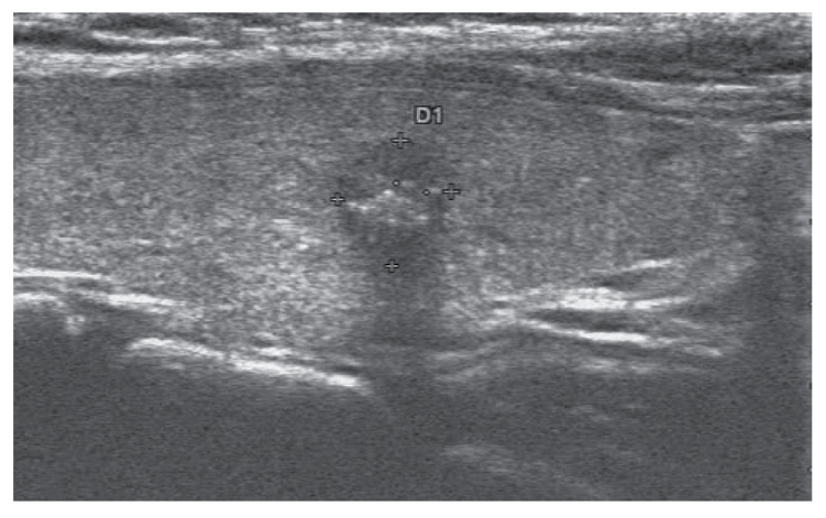
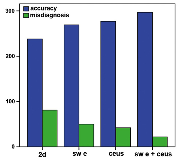
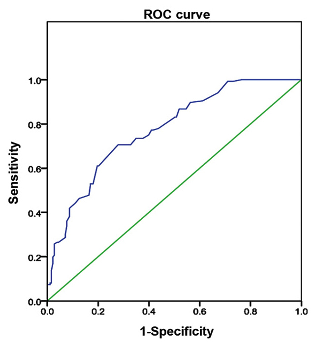
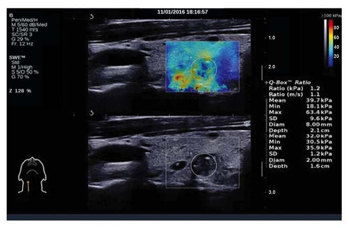
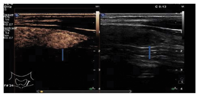
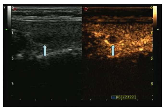
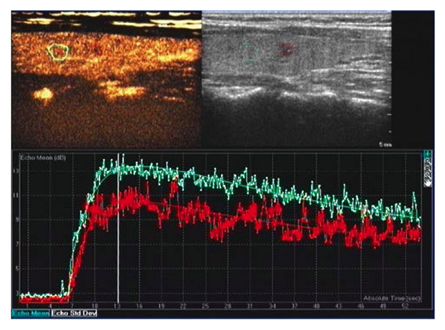
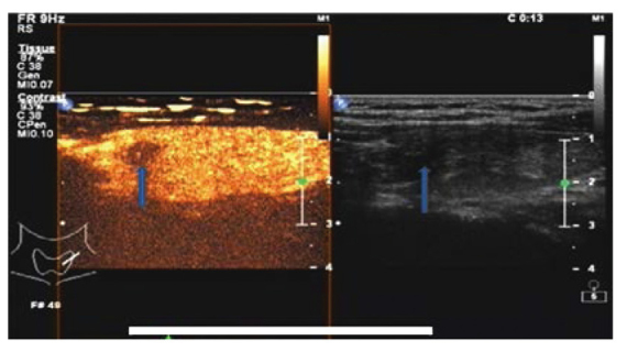
The aim of the present study was to evaluate the value of shear wave elastography (SWE) and contrast-enhanced ultrasonography (CEUS) in the diagnosis of thyroid malignant nodules. A total of 253 patients with 319 thyroid nodules were subjected to two-dimensional ultrasound (2DUS) and CEUS examinations prior to thyroidectomy between March, 2014 and December, 2015. Young's modulus value for each nodule on 2DUS and CEUS images were recorded. The sensitivity, specificity and accuracy of 2DUS, SWE and CEUS in the diagnosis of thyroid malignant nodules were assessed. The results demonstrated that, of the 319 nodules that were pathologically confirmed, 183 were malignant and 136 were benign. The area under the receiver operating characteristic curve as a result of SWE diagnosis was 0.77. When the threshold of the Young's modulus value was ≥27.65 kPa in the diagnosis of malignant thyroid nodules, SWE exhibited a sensitivity of 84.55% (115/136), a specificity of 84.15% (154/183) and an accuracy of 84.32% (269/319). US contrast imaging of malignant thyroid nodules revealed a major tendency for early hypoenhancement and hypoenhancement. CEUS exhibited a sensitivity of 87.5% (119/136), a specificity of 86.33% (158/183) and an accuracy of 86.83% (277/319) in the diagnosis of malignant thyroid nodules. Compared with 2DUS, SWE, CEUS and their combined use exhibited statistically significant differences in the diagnosis of thyroid malignant nodules in terms of sensitivity, specificity and accuracy (χ=9.220,15.310 and 40.296, respectively; P=0.000); SWE or CEUS did not differ significantly in the diagnosis of thyroid malignant nodules in terms of sensitivity, specificity or accuracy (χ=0.737;P=0.542); Compared with the use of SWE or CEUS alone, their combination exhibited statistically significant differences in the diagnosis of malignant thyroid nodules in terms of sensitivity, specificity and accuracy (χ=12.264 and 6.939, respectively; P=0.000,0.005). In conclusion, the high accuracy of the combined use of SWE and CEUS in the diagnosis of malignant thyroid nodules is of great clinical value.

摘要

本研究旨在评估剪切波弹性成像(SWE)和超声造影(CEUS)在甲状腺恶性结节诊断中的价值。2014年3月至2015年12月期间,共有253例患有319个甲状腺结节的患者在甲状腺切除术前接受了二维超声(2DUS)和CEUS检查。记录2DUS和CEUS图像上每个结节的杨氏模量值。评估2DUS、SWE和CEUS在甲状腺恶性结节诊断中的敏感性、特异性和准确性。结果表明,在319个经病理证实的结节中,183个为恶性,136个为良性。SWE诊断的受试者操作特征曲线下面积为0.77。在诊断甲状腺恶性结节时,当杨氏模量值阈值≥27.65 kPa时,SWE的敏感性为84.55%(115/136),特异性为84.15%(154/183),准确性为84.32%(269/319)。甲状腺恶性结节的超声造影成像显示早期低增强和低增强的主要趋势。CEUS在甲状腺恶性结节诊断中的敏感性为87.5%(119/136),特异性为86.33%(158/183),准确性为86.83%(277/319)。与2DUS相比,SWE、CEUS及其联合使用在甲状腺恶性结节诊断的敏感性、特异性和准确性方面存在统计学显著差异(χ分别为9.220、15.310和40.296;P = 0.000);SWE或CEUS在甲状腺恶性结节诊断的敏感性、特异性或准确性方面无显著差异(χ = 0.737;P = 0.542);与单独使用SWE或CEUS相比,它们的联合使用在甲状腺恶性结节诊断的敏感性、特异性和准确性方面存在统计学显著差异(χ分别为12.264和6.939;P = 0.000、0.005)。总之,SWE和CEUS联合使用在甲状腺恶性结节诊断中的高准确性具有重要的临床价值。

https://cdn.ncbi.nlm.nih.gov/pmc/blobs/8856/5228169/4d73ed75024f/mco-05-06-0724-g07.jpg
https://cdn.ncbi.nlm.nih.gov/pmc/blobs/8856/5228169/72c3739e947f/mco-05-06-0724-g00.jpg
https://cdn.ncbi.nlm.nih.gov/pmc/blobs/8856/5228169/66095d8ce795/mco-05-06-0724-g01.jpg
https://cdn.ncbi.nlm.nih.gov/pmc/blobs/8856/5228169/a09cef6051d7/mco-05-06-0724-g02.jpg
https://cdn.ncbi.nlm.nih.gov/pmc/blobs/8856/5228169/d47219e6c48c/mco-05-06-0724-g03.jpg
https://cdn.ncbi.nlm.nih.gov/pmc/blobs/8856/5228169/90162ad45c51/mco-05-06-0724-g04.jpg
https://cdn.ncbi.nlm.nih.gov/pmc/blobs/8856/5228169/94b6c2b52224/mco-05-06-0724-g05.jpg
https://cdn.ncbi.nlm.nih.gov/pmc/blobs/8856/5228169/8efe12fa45e1/mco-05-06-0724-g06.jpg
https://cdn.ncbi.nlm.nih.gov/pmc/blobs/8856/5228169/4d73ed75024f/mco-05-06-0724-g07.jpg

相似文献

1
Shear wave elastography and contrast-enhanced ultrasonography in the diagnosis of thyroid malignant nodules.剪切波弹性成像和超声造影在甲状腺恶性结节诊断中的应用
Mol Clin Oncol. 2016 Dec;5(6):724-730. doi: 10.3892/mco.2016.1053. Epub 2016 Oct 19.
2
Contrast-enhanced ultrasound and shear wave elastography in the diagnosis of ACR TI-RADS 4 and 5 category thyroid nodules coexisting with Hashimoto's thyroiditis.超声造影及剪切波弹性成像在诊断合并桥本甲状腺炎的ACR TI-RADS 4类和5类甲状腺结节中的应用
Front Oncol. 2023 Jan 11;12:1022305. doi: 10.3389/fonc.2022.1022305. eCollection 2022.
3
Application of ultrasonic shear wave elastography and contrast-enhanced ultrasound in the differential diagnosis of patients with benign and malignant thyroid lesions.超声剪切波弹性成像与超声造影在甲状腺良恶性病变患者鉴别诊断中的应用
Gland Surg. 2020 Dec;9(6):2136-2143. doi: 10.21037/gs-20-819.
4
Incremental diagnostic value of shear wave elastography combined with contrast-enhanced ultrasound in TI-RADS category 4a and 4b nodules.剪切波弹性成像联合超声造影在TI-RADS 4a和4b类结节中的增量诊断价值
J Med Ultrason (2001). 2020 Jul;47(3):453-462. doi: 10.1007/s10396-020-01016-8. Epub 2020 Apr 18.
5
[The diagnostic value of the maximum value of Young's modulus of shear-wave elastography and ACR TI-RADS for thyroid nodules].[剪切波弹性成像杨氏模量最大值及ACR TI-RADS对甲状腺结节的诊断价值]
Lin Chuang Er Bi Yan Hou Tou Jing Wai Ke Za Zhi. 2019 Aug;33(8):764-767. doi: 10.13201/j.issn.1001-1781.2019.08.020.
6
Significance of contrast-enhanced ultrasonography in differential diagnosis of thyroid nodules.超声造影在甲状腺结节鉴别诊断中的意义
Medicine (Baltimore). 2018 Oct;97(40):e12688. doi: 10.1097/MD.0000000000012688.
7
[Value of thyroid imaging reporting and data system and shear wave elastography for diagnosis of thyroid microcarcinoma].[甲状腺影像报告和数据系统及剪切波弹性成像在甲状腺微小癌诊断中的价值]
Lin Chuang Er Bi Yan Hou Tou Jing Wai Ke Za Zhi. 2016 Jan;30(2):118-22.
8
Clinical Study of the Prediction of Malignancy in Thyroid Nodules: Modified Score versus 2017 American College of Radiology's Thyroid Imaging Reporting and Data System Ultrasound Lexicon.甲状腺结节良恶性预测的临床研究:改良评分与 2017 年美国放射学会甲状腺影像报告和数据系统超声词汇的比较。
Ultrasound Med Biol. 2019 Jul;45(7):1627-1637. doi: 10.1016/j.ultrasmedbio.2019.03.014. Epub 2019 May 4.
9
Diagnostic performance of C-TIRADS combined with SWE for the diagnosis of thyroid nodules.C-TIRADS 联合超声弹性成像技术对甲状腺结节的诊断性能。
Front Endocrinol (Lausanne). 2022 Sep 6;13:939303. doi: 10.3389/fendo.2022.939303. eCollection 2022.
10
Combination of contrast-enhanced ultrasound and strain elastography to assess cytologically non-diagnostic thyroid nodules.超声造影与应变弹性成像联合评估细胞学检查未明确诊断的甲状腺结节
Oncol Lett. 2019 Dec;18(6):6845-6851. doi: 10.3892/ol.2019.11058. Epub 2019 Nov 5.

引用本文的文献

1
Diagnostic value of combined ultrasound contrast and elastography for differentiating benign and malignant thyroid nodules: a meta-analysis.超声造影联合弹性成像技术鉴别良恶性甲状腺结节的诊断价值:Meta 分析。
Sci Rep. 2024 Jun 1;14(1):12605. doi: 10.1038/s41598-024-63420-1.
2
Incorporation of CEUS and SWE parameters into a multivariate logistic regression model for the differential diagnosis of benign and malignant TI-RADS 4 thyroid nodules.将 CEUS 和 SWE 参数纳入多元逻辑回归模型,用于鉴别诊断 TI-RADS 4 级甲状腺良恶性结节。
Endocrine. 2024 Mar;83(3):691-699. doi: 10.1007/s12020-023-03524-2. Epub 2023 Oct 27.
3

本文引用的文献

1
Role of contrast-enhanced ultrasound (CEUS) in the diagnosis of endometrial pathology.超声造影(CEUS)在子宫内膜病变诊断中的作用。
Clujul Med. 2015;88(4):433-7. doi: 10.15386/cjmed-499. Epub 2015 Nov 15.
2
Contrast-enhanced ultrasound for evaluation of renal trauma during acute hemorrhagic shock: a canine model.超声造影评估急性失血性休克期间的肾损伤:犬模型
J Med Ultrason (2001). 2015 Apr;42(2):199-205. doi: 10.1007/s10396-014-0601-5. Epub 2014 Dec 19.
3
Conventional US, elastography, and contrast enhanced US features of papillary thyroid microcarcinoma predict central compartment lymph node metastases.
Quantification of Thyroid Viscosity in Healthy Subjects Using Ultrasound Shear Wave Dispersion (Viscosity PLUS).
使用超声剪切波频散技术(Viscosity PLUS)对健康受试者甲状腺黏度进行定量分析。
Diagnostics (Basel). 2022 Sep 9;12(9):2194. doi: 10.3390/diagnostics12092194.
4
Application of Multiple Ultrasonic Techniques in the Diagnosis of Prostate Cancer.多种超声技术在前列腺癌诊断中的应用
Front Oncol. 2022 Jun 27;12:905087. doi: 10.3389/fonc.2022.905087. eCollection 2022.
5
US-Elastography With Different Techniques for Thyroid Nodule Characterization: Systematic Review and Meta-analysis.采用不同技术的超声弹性成像对甲状腺结节的特征分析:系统评价与荟萃分析
Front Oncol. 2022 Mar 16;12:845549. doi: 10.3389/fonc.2022.845549. eCollection 2022.
6
SuperSonic shear imaging for the differentiation between benign and malignant thyroid nodules: a meta-analysis.超声剪切波弹性成像在鉴别甲状腺良恶性结节中的应用:一项荟萃分析
J Endocrinol Invest. 2022 Jul;45(7):1327-1339. doi: 10.1007/s40618-022-01765-y. Epub 2022 Mar 1.
7
Performance of Contrast-Enhanced Ultrasound in Thyroid Nodules: Review of Current State and Future Perspectives.超声造影在甲状腺结节中的应用:现状与未来展望
Cancers (Basel). 2021 Oct 30;13(21):5469. doi: 10.3390/cancers13215469.
8
Evaluation of thyroid nodules by shear wave elastography: a review of current knowledge.剪切波弹性成像评估甲状腺结节:当前知识综述。
J Endocrinol Invest. 2021 Oct;44(10):2043-2056. doi: 10.1007/s40618-021-01570-z. Epub 2021 Apr 16.
9
Multimode ultrasonic technique is recommended for the differential diagnosis of thyroid cancer.多模式超声技术被推荐用于甲状腺癌的鉴别诊断。
PeerJ. 2020 May 4;8:e9112. doi: 10.7717/peerj.9112. eCollection 2020.
10
Contrast-enhanced ultrasound for the differential diagnosis of thyroid nodules: An updated meta-analysis with comprehensive heterogeneity analysis.超声造影对甲状腺结节的鉴别诊断:一项包含全面异质性分析的更新荟萃分析。
PLoS One. 2020 Apr 20;15(4):e0231775. doi: 10.1371/journal.pone.0231775. eCollection 2020.
甲状腺微小乳头状癌的传统超声、弹性成像及超声造影特征可预测中央区淋巴结转移。
Sci Rep. 2015 Jan 13;5:7748. doi: 10.1038/srep07748.
4
Contrast-enhanced sonography of thyroid nodules.甲状腺结节的超声造影检查
J Clin Ultrasound. 2015 Mar;43(3):153-6. doi: 10.1002/jcu.22240. Epub 2014 Oct 18.
5
Shear wave elastography for detection of prostate cancer: a preliminary study.剪切波弹性成像用于前列腺癌检测的初步研究。
Korean J Radiol. 2014 May-Jun;15(3):346-55. doi: 10.3348/kjr.2014.15.3.346. Epub 2014 Apr 29.
6
Diagnostic performances of various gray-scale, color Doppler, and contrast-enhanced ultrasonography findings in predicting malignant thyroid nodules.各种灰阶、彩色多普勒和超声造影超声表现对预测甲状腺恶性结节的诊断性能。
Thyroid. 2014 Feb;24(2):355-63. doi: 10.1089/thy.2013.0150. Epub 2013 Nov 14.
7
Prospective comparative evaluation of quantitative-elastosonography (Q-elastography) and contrast-enhanced ultrasound for the evaluation of thyroid nodules: preliminary experience.前瞻性定量超声弹性成像(Q 弹性成像)与超声造影对甲状腺结节评估的对比研究:初步经验。
Eur J Radiol. 2013 Nov;82(11):1892-8. doi: 10.1016/j.ejrad.2013.07.005. Epub 2013 Aug 6.
8
Number of central lymph node metastasis for predicting lateral lymph node metastasis in papillary thyroid microcarcinoma.中央区淋巴结转移数目预测甲状腺微小乳头状癌侧颈区淋巴结转移的研究
Head Neck. 2014 Jan;36(1):101-6. doi: 10.1002/hed.23270. Epub 2013 Jul 30.
9
The sonographic appearance of benign and malignant thyroid diseases and their histopathology correlate: demystifying the thyroid nodule.甲状腺良恶性疾病的超声表现及其组织病理学相关性:解读甲状腺结节
Ultrasound Q. 2013 Sep;29(3):161-78. doi: 10.1097/RUQ.0b013e31829a573e.
10
Comparative study of thyroid puncture biopsy guided by contrast-enhanced ultrasonography and conventional ultrasound.超声造影引导与传统超声引导下甲状腺穿刺活检的对比研究
Exp Ther Med. 2013 May;5(5):1381-1384. doi: 10.3892/etm.2013.1016. Epub 2013 Mar 15.