Kale Alok, Joshi Priscilla, Kelkar A B
Department of Radiodiagnosis and Imaging, Bharati Hospital and Research Center, Pune, Maharashtra, India.
Indian J Radiol Imaging. 2016 Oct-Dec;26(4):487-492. doi: 10.4103/0971-3026.195795.
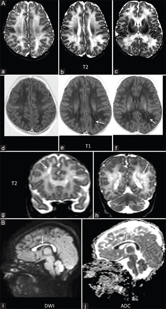
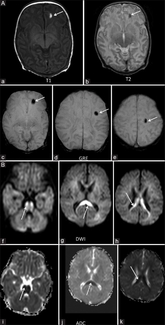
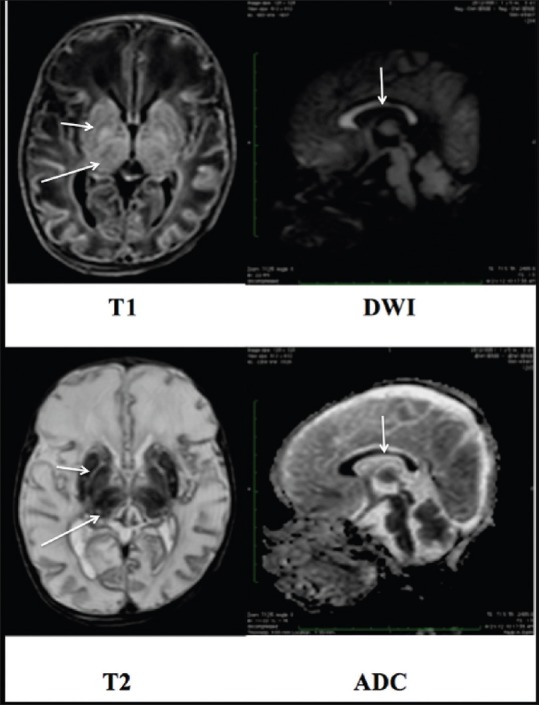
Restricted diffusion within the splenium of the corpus callosum has been described by other authors in various conditions, however, restricted diffusion in the entire corpus callosum or isolated involvement of the splenium, genu, or body has been infrequently reported on magnetic resonance imaging (MRI) in neonatal hypoxic-ischemic encephalopathy. We report a series of cases showing different patterns of involvement.
Perinatal imaging with MRI including diffusion-weighted imaging was performed in 40 neonates with hypoxic-ischemic encephalopathy, including 11 premature neonates. Sixteen out of 40 patients demonstrated restricted diffusion within the corpus callosum. Out of 16 patients, 9 showed restricted diffusion in the entire corpus callosum, 4 had isolated splenium involvement, 2 had body and splenium signal abnormality, and 1 showed diffusion restriction only in the genu.
Changes in the corpus callosum were also associated with more severe clinical presentation of encephalopathy. Restricted diffusion within the corpus callosum in infants with hypoxic-ischemic encephalopathy is often associated with extensive brain injury and appears to be an early neuroradiologic marker of adverse neurologic outcome.
其他作者已在多种情况下描述了胼胝体压部的扩散受限,然而,在新生儿缺氧缺血性脑病的磁共振成像(MRI)中,胼胝体整体扩散受限或仅累及压部、膝部或体部的情况鲜有报道。我们报告了一系列显示不同受累模式的病例。
对40例缺氧缺血性脑病新生儿进行了包括扩散加权成像在内的围产期MRI检查,其中包括11例早产儿。40例患者中有16例在胼胝体内出现扩散受限。在这16例患者中,9例在整个胼胝体出现扩散受限,4例仅累及压部,2例体部和压部信号异常,1例仅在膝部出现扩散受限。
胼胝体的变化也与更严重的脑病临床表现相关。缺氧缺血性脑病婴儿胼胝体内的扩散受限通常与广泛的脑损伤有关,似乎是不良神经预后的早期神经放射学标志。