Van 't Hoff Institute for Molecular Sciences, University of Amsterdam, Science Park 904, 1098 XH Amsterdam, The Netherlands.
Nanoscale Biophysics Group, FOM Institute AMOLF, Science Park 104, 1098 XG Amsterdam, The Netherlands.
Sci Rep. 2017 Jan 23;7:41051. doi: 10.1038/srep41051.
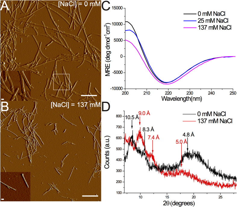
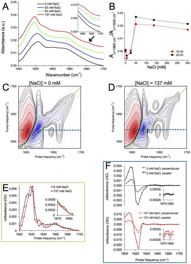
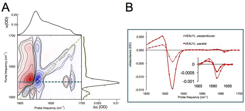
The aggregation of the intrinsically disordered protein alpha-synuclein (αS) into amyloid fibrils is thought to play a central role in the pathology of Parkinson's disease. Using a combination of techniques (AFM, UV-CD, XRD, and amide-I 1D- and 2D-IR spectroscopy) we show that the structure of αS fibrils varies as a function of ionic strength: fibrils aggregated in low ionic-strength buffers ([NaCl] ≤ 25 mM) have a significantly different structure than fibrils grown in higher ionic-strength buffers. The observations for fibrils aggregated in low-salt buffers are consistent with an extended conformation of αS molecules, forming hydrogen-bonded intermolecular β-sheets that are loosely packed in a parallel fashion. For fibrils aggregated in high-salt buffers (including those prepared in buffers with a physiological salt concentration) the measurements are consistent with αS molecules in a more tightly-packed, antiparallel intramolecular conformation, and suggest a structure characterized by two twisting stacks of approximately five hydrogen-bonded intermolecular β-sheets each. We find evidence that the high-frequency peak in the amide-I spectrum of αS fibrils involves a normal mode that differs fundamentally from the canonical high-frequency antiparallel β-sheet mode. The high sensitivity of the fibril structure to the ionic strength might form the basis of differences in αS-related pathologies.
α-突触核蛋白(αS)的无规卷曲蛋白聚集形成淀粉样纤维被认为在帕金森病的病理中起核心作用。我们使用多种技术(原子力显微镜、紫外圆二色性、X 射线衍射和酰胺 I 的一维和二维红外光谱)表明,αS 纤维的结构随离子强度而变化:在低离子强度缓冲液([NaCl]≤25 mM)中聚集的纤维的结构与在较高离子强度缓冲液中生长的纤维有显著差异。在低盐缓冲液中聚集的纤维的观察结果与αS 分子的伸展构象一致,形成氢键相互作用的分子间β-片层,以平行方式松散堆积。对于在高盐缓冲液中聚集的纤维(包括在生理盐浓度的缓冲液中制备的纤维),测量结果与更紧密堆积的、反平行的分子内构象一致,并表明结构的特征是每个扭转堆叠有大约五个氢键相互作用的分子间β-片层。我们发现证据表明,αS 纤维的酰胺 I 光谱中的高频峰涉及一种与典型的高频反平行β-片层模式在根本上不同的本征模式。纤维结构对离子强度的高度敏感性可能是与 αS 相关的病理学差异的基础。