Sarkari Bahador, Fatemie Sfedan Asieh, Moshfe Abdolali, Abdolahi Khabisi Samaneh, Savardashtaki Amir, Hosseini Farshad, Shahbazi Ardavan
Basic Sciences in Infectious Diseases Research Center, Shiraz University of Medical Sciences, Shiraz, Iran.
Dept. of Parasitology and Mycology, School of Medicine, Shiraz University of Medical Sciences, Shiraz, Iran.
Iran J Parasitol. 2016 Oct-Dec;11(4):585-590.
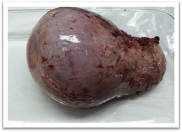
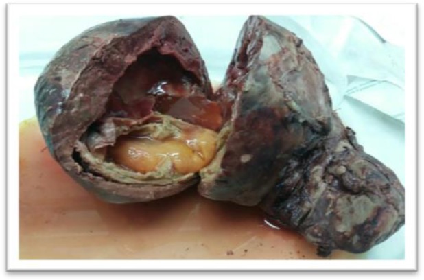
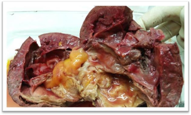
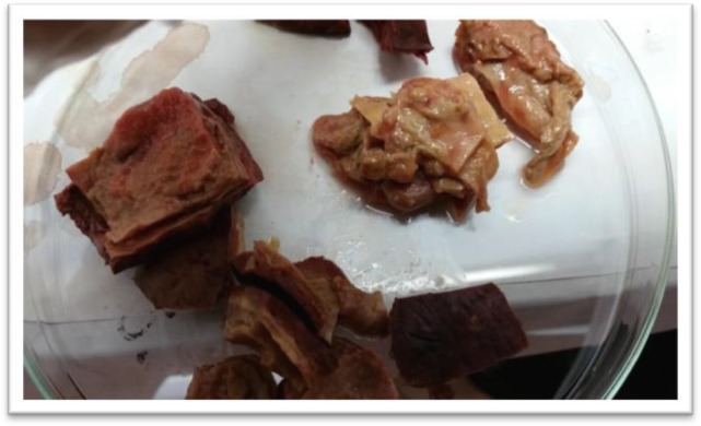
Spleen is an unusual location for hydatid cyst. Here we report a case of primary splenic hydatid cyst in a 41-yr-old Iranian woman from Yasuj, southwest of Iran. The patient had been admitted to Shahid Beheshti Hospital because of abdominal pain. Abdominal sonography revealed a hypoechoic lesion of 150 X 130 mm in the spleen, suggestive of hydatid cyst. Splenectomy was performed for the patient and surgical interventions revealed a hydatid cyst occupying most of splenic parenchyma. She was discharged on the 5 day of her operation. Postoperative diagnosis and confirmation of hydatid cyst was done by histopathological, molecular and serological approaches. Histopathological evaluation revealed the classical laminated layer of hydatid cyst. DNA was extracted from a part of cyst and PCR amplified. Sequencing and analysis of PCR product revealed that the isolate has the most similarity with G1 strain of . Patient's serum was positive for IgG anti-hydatid cyst antibodies, using antigen-B ELISA.
脾脏是包虫囊肿的罕见发病部位。在此,我们报告一例来自伊朗西南部亚苏季的41岁伊朗女性原发性脾包虫囊肿病例。该患者因腹痛入住沙希德·贝赫什提医院。腹部超声检查发现脾脏有一个150×130毫米的低回声病变,提示为包虫囊肿。为该患者实施了脾切除术,手术发现一个包虫囊肿占据了大部分脾实质。她在术后第5天出院。通过组织病理学、分子学和血清学方法对包虫囊肿进行了术后诊断和确认。组织病理学评估显示了包虫囊肿典型的板层结构。从囊肿的一部分提取了DNA并进行PCR扩增。PCR产物的测序和分析表明,分离株与 的G1菌株最为相似。使用抗原-B ELISA检测,患者血清中抗包虫囊肿IgG抗体呈阳性。