• 文献检索
  • 文档翻译
  • 深度研究
  • 学术资讯
  • Suppr Zotero 插件Zotero 插件
  • 邀请有礼
  • 套餐&价格
  • 历史记录
应用&插件
Suppr Zotero 插件Zotero 插件浏览器插件Mac 客户端Windows 客户端微信小程序
定价
高级版会员购买积分包购买API积分包
服务
文献检索文档翻译深度研究API 文档MCP 服务
关于我们
关于 Suppr公司介绍联系我们用户协议隐私条款
关注我们

Suppr 超能文献

核心技术专利:CN118964589B侵权必究
粤ICP备2023148730 号-1Suppr @ 2026

文献检索

告别复杂PubMed语法,用中文像聊天一样搜索,搜遍4000万医学文献。AI智能推荐,让科研检索更轻松。

立即免费搜索

文件翻译

保留排版,准确专业,支持PDF/Word/PPT等文件格式,支持 12+语言互译。

免费翻译文档

深度研究

AI帮你快速写综述,25分钟生成高质量综述,智能提取关键信息,辅助科研写作。

立即免费体验

静息态功能磁共振成像在亚急性缺血性卒中的价值:与动态磁敏感对比增强灌注磁共振成像的比较。

The value of resting-state functional MRI in subacute ischemic stroke: comparison with dynamic susceptibility contrast-enhanced perfusion MRI.

机构信息

Department of Radiology, The Affiliated Drum Tower Hospital of Nanjing University Medical School, University of Nanjing, Nanjing, China.

Department of Neurology, The Affiliated Drum Tower Hospital of Nanjing University Medical School, Nanjing, China.

出版信息

Sci Rep. 2017 Jan 31;7:41586. doi: 10.1038/srep41586.

DOI:10.1038/srep41586
PMID:28139701
原文链接:https://pmc.ncbi.nlm.nih.gov/articles/PMC5282488/
Abstract

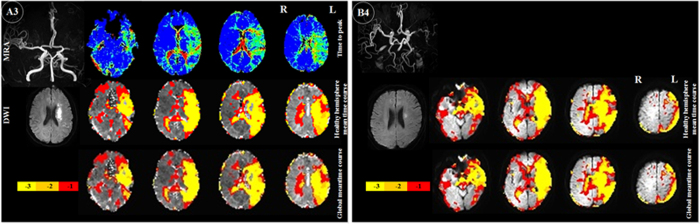
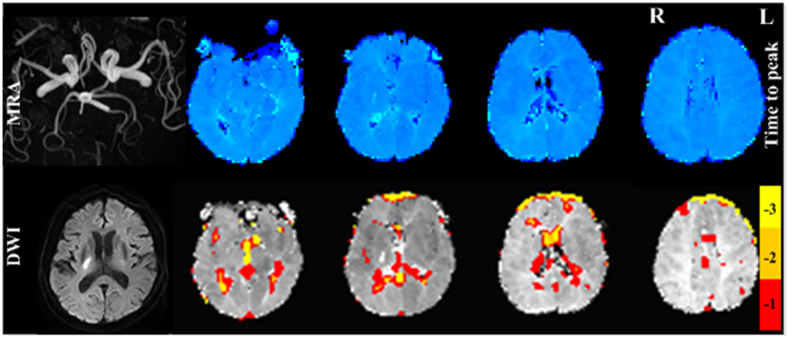
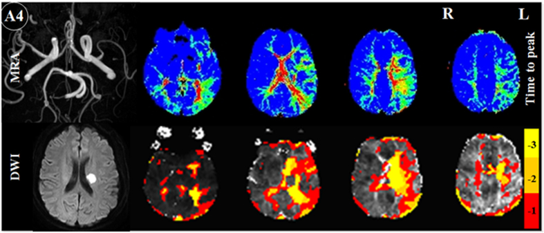
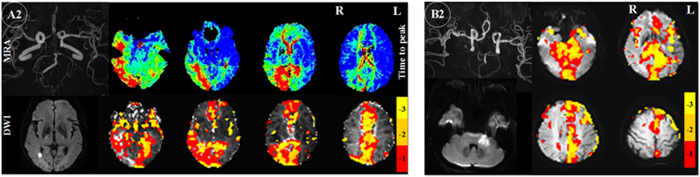
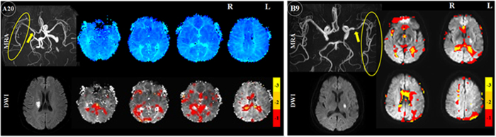
To evaluate the potential clinical value of the time-shift analysis (TSA) approach for resting-state fMRI (rs-fMRI) blood oxygenation level-dependent (BOLD) data in detecting hypoperfusion of subacute stroke patients through comparison with dynamic susceptibility contrast perfusion weighted imaging (DSC-PWI). Forty patients with subacute stroke (3-14 days after neurological symptom onset) underwent MRI examination. Cohort A: 31 patients had MRA, DSC-PWI and BOLD data. Cohort B: 9 patients had BOLD and MRA data. The time delay between the BOLD time course in each voxel and the mean signal of global and contralateral hemisphere was calculated using TSA. Time to peak (TTP) was employed to detect hypoperfusion. Among cohort A, 14 patients who had intracranial large-vessel occlusion/stenosis with sparse collaterals showed hypoperfusion by both of the two approaches, one with abundant collaterals showed neither TTP nor TSA time delay. The remaining 16 patients without obvious MRA lesions showed neither TTP nor TSA time delay. Among cohort B, eight patients showed time delay areas. The TSA approach was a promising alternative to DSC-PWI for detecting hypoperfusion in subacute stroke patients who had obvious MRA lesions with sparse collaterals, those with abundant collaterals would keep intact local perfusion.

摘要

为了评估时间漂移分析(TSA)方法在检测亚急性脑卒中患者低灌注中的潜在临床价值,我们通过与动态磁敏感对比灌注加权成像(DSC-PWI)进行比较,对亚急性脑卒中患者(发病后 3-14 天)的静息态功能磁共振成像(rs-fMRI)血氧水平依赖(BOLD)数据进行了评估。40 例亚急性脑卒中患者接受了 MRI 检查。队列 A:31 例患者有 MRA、DSC-PWI 和 BOLD 数据。队列 B:9 例患者有 BOLD 和 MRA 数据。使用 TSA 计算每个体素的 BOLD 时程与全脑和对侧半球平均信号之间的时间延迟。采用达峰时间(TTP)来检测低灌注。在队列 A 中,14 例颅内大血管闭塞/狭窄伴稀疏侧支循环的患者,两种方法均显示低灌注,其中 1 例侧支循环丰富的患者未出现 TTP 或 TSA 时间延迟。其余 16 例无明显 MRA 病变的患者均未出现 TTP 或 TSA 时间延迟。在队列 B 中,8 例患者出现时间延迟区域。在明显有稀疏侧支循环的 MRA 病变且伴丰富侧支循环的亚急性脑卒中患者中,TSA 方法是一种很有前途的 DSC-PWI 替代方法,可以检测低灌注,而局部灌注保持完整。

https://cdn.ncbi.nlm.nih.gov/pmc/blobs/f136/5282488/18b07a5fc2d7/srep41586-f5.jpg
https://cdn.ncbi.nlm.nih.gov/pmc/blobs/f136/5282488/386a98f65d2b/srep41586-f1.jpg
https://cdn.ncbi.nlm.nih.gov/pmc/blobs/f136/5282488/ba67b06616d0/srep41586-f2.jpg
https://cdn.ncbi.nlm.nih.gov/pmc/blobs/f136/5282488/cf71046f1db8/srep41586-f3.jpg
https://cdn.ncbi.nlm.nih.gov/pmc/blobs/f136/5282488/76c5ffe43496/srep41586-f4.jpg
https://cdn.ncbi.nlm.nih.gov/pmc/blobs/f136/5282488/18b07a5fc2d7/srep41586-f5.jpg

相似文献

1
The value of resting-state functional MRI in subacute ischemic stroke: comparison with dynamic susceptibility contrast-enhanced perfusion MRI.静息态功能磁共振成像在亚急性缺血性卒中的价值:与动态磁敏感对比增强灌注磁共振成像的比较。
Sci Rep. 2017 Jan 31;7:41586. doi: 10.1038/srep41586.
2
Cerebral hemodynamic impairment: assessment with resting-state functional MR imaging.脑血流动力学障碍:静息态功能磁共振成像评估。
Radiology. 2014 Feb;270(2):548-55. doi: 10.1148/radiol.13130982. Epub 2013 Oct 28.
3
One-step analysis of brain perfusion and function for acute stroke patients after reperfusion: A resting-state fMRI study.急性卒中再灌注后患者脑灌注与功能的一步分析:静息态 fMRI 研究。
J Magn Reson Imaging. 2019 Jul;50(1):221-229. doi: 10.1002/jmri.26571. Epub 2018 Dec 19.
4
Acute stroke imaging: feasibility and value of MR angiography with high spatial and temporal resolution for vessel assessment and perfusion analysis in patients with wake-up stroke.急性卒中成像:高空间和时间分辨率磁共振血管造影在醒后卒中患者血管评估和灌注分析中的可行性及价值
Acad Radiol. 2015 Apr;22(4):413-22. doi: 10.1016/j.acra.2014.11.013. Epub 2015 Jan 15.
5
Relationship Between Changes in the Temporal Dynamics of the Blood-Oxygen-Level-Dependent Signal and Hypoperfusion in Acute Ischemic Stroke.急性缺血性卒中患者血氧水平依赖信号的时间动态变化与灌注不足之间的关系
Stroke. 2017 Apr;48(4):925-931. doi: 10.1161/STROKEAHA.116.015566. Epub 2017 Mar 8.
6
Perfusion Maps Acquired From Dynamic Angiography MRI Using Deep Learning Approaches.使用深度学习方法从动态血管造影磁共振成像中获取的灌注图。
J Magn Reson Imaging. 2023 Feb;57(2):456-469. doi: 10.1002/jmri.28315. Epub 2022 Jun 21.
7
Dynamic susceptibility contrast perfusion MRI identifies persistent vessel pathology in acute pontine stroke.动态磁敏感对比灌注 MRI 可识别急性脑桥卒中的持续性血管病变。
Cerebrovasc Dis. 2010;29(4):389-94. doi: 10.1159/000286341. Epub 2010 Feb 19.
8
Toward fully automated processing of dynamic susceptibility contrast perfusion MRI for acute ischemic cerebral stroke.实现急性缺血性脑卒中动态磁敏感对比灌注 MRI 全自动处理。
Comput Methods Programs Biomed. 2010 May;98(2):204-13. doi: 10.1016/j.cmpb.2009.12.005. Epub 2010 Jan 8.
9
Iron Sucrose as MRI Contrast Agent in Ischemic Stroke Model.蔗糖铁作为缺血性中风模型中的磁共振成像造影剂
J Magn Reson Imaging. 2020 Sep;52(3):836-849. doi: 10.1002/jmri.27109. Epub 2020 Feb 29.
10
Non-invasive monitoring of longitudinal changes in cerebral hemodynamics in acute ischemic stroke using BOLD signal delay.使用 BOLD 信号延迟无创监测急性缺血性脑卒中患者大脑血液动力学的纵向变化。
J Cereb Blood Flow Metab. 2020 Jan;40(1):23-34. doi: 10.1177/0271678X18803951. Epub 2018 Oct 18.

引用本文的文献

1
Deep-learning-enabled brain hemodynamic mapping using resting-state fMRI.使用静息态功能磁共振成像的深度学习脑血流动力学图谱绘制
NPJ Digit Med. 2023 Jun 21;6(1):116. doi: 10.1038/s41746-023-00859-y.
2
Hemodynamic timing in resting-state and breathing-task BOLD fMRI.静息态和呼吸任务 BOLD fMRI 的血流动力学时程。
Neuroimage. 2023 Jul 1;274:120120. doi: 10.1016/j.neuroimage.2023.120120. Epub 2023 Apr 16.
3
Reproducibility of cerebral perfusion measurements using BOLD delay.使用 BOLD 延迟测量脑灌注的可重复性。

本文引用的文献

1
Cerebral Blood Volume ASPECTS Is the Best Predictor of Clinical Outcome in Acute Ischemic Stroke: A Retrospective, Combined Semi-Quantitative and Quantitative Assessment.脑血容量ASPECTS是急性缺血性卒中临床结局的最佳预测指标:一项回顾性、半定量与定量相结合的评估研究
PLoS One. 2016 Jan 29;11(1):e0147910. doi: 10.1371/journal.pone.0147910. eCollection 2016.
2
The effects of hemodynamic lag on functional connectivity and behavior after stroke.血流动力学延迟对中风后功能连接和行为的影响。
J Cereb Blood Flow Metab. 2016 Dec;36(12):2162-2176. doi: 10.1177/0271678X15614846. Epub 2015 Nov 4.
3
Intracranial Gadolinium Deposition after Contrast-enhanced MR Imaging.
Hum Brain Mapp. 2023 May;44(7):2778-2789. doi: 10.1002/hbm.26244. Epub 2023 Feb 25.
4
RP-Rs-fMRIomics as a Novel Imaging Analysis Strategy to Empower Diagnosis of Brain Gliomas.RP-Rs-fMRIomics作为一种新型成像分析策略助力脑胶质瘤诊断
Cancers (Basel). 2022 Jun 7;14(12):2818. doi: 10.3390/cancers14122818.
5
Artificially-reconstructed brain images with stroke lesions from non-imaging data: modeling in categorized patients based on lesion occurrence and sparsity.基于病灶发生和稀疏性对分类患者进行建模:利用非影像数据构建具有中风病灶的人工重建脑图像。
Sci Rep. 2022 Jun 16;12(1):10116. doi: 10.1038/s41598-022-14249-z.
6
Coupling between cerebrovascular oscillations and CSF flow fluctuations during wakefulness: An fMRI study.清醒状态下脑血流波动与 CSF 流动波动的耦合:一项 fMRI 研究。
J Cereb Blood Flow Metab. 2022 Jun;42(6):1091-1103. doi: 10.1177/0271678X221074639. Epub 2022 Jan 16.
7
Effects of Thresholding on Voxel-Wise Correspondence of Breath-Hold and Resting-State Maps of Cerebrovascular Reactivity.阈值处理对脑血管反应性屏气和静息态图谱体素对应关系的影响。
Front Neurosci. 2021 Aug 24;15:654957. doi: 10.3389/fnins.2021.654957. eCollection 2021.
8
A novel approach for assessing hypoperfusion in stroke using spatial independent component analysis of resting-state fMRI.利用静息态 fMRI 的空间独立成分分析评估卒中低灌注的新方法。
Hum Brain Mapp. 2021 Nov;42(16):5204-5216. doi: 10.1002/hbm.25610. Epub 2021 Jul 29.
9
A practical modification to a resting state fMRI protocol for improved characterization of cerebrovascular function.一种改进静息态 fMRI 协议以改善脑血管功能表征的实用方法。
Neuroimage. 2021 Oct 1;239:118306. doi: 10.1016/j.neuroimage.2021.118306. Epub 2021 Jun 24.
10
Vector Phase Analysis Approach for Sleep Stage Classification: A Functional Near-Infrared Spectroscopy-Based Passive Brain-Computer Interface.用于睡眠阶段分类的矢量相位分析方法:一种基于功能近红外光谱的被动式脑机接口
Front Hum Neurosci. 2021 Apr 30;15:658444. doi: 10.3389/fnhum.2021.658444. eCollection 2021.
颅内钆沉积与对比增强磁共振成像后。
Radiology. 2015 Jun;275(3):772-82. doi: 10.1148/radiol.15150025. Epub 2015 Mar 5.
4
Endovascular therapy for ischemic stroke with perfusion-imaging selection.血管内治疗缺血性卒中的灌注成像选择。
N Engl J Med. 2015 Mar 12;372(11):1009-18. doi: 10.1056/NEJMoa1414792. Epub 2015 Feb 11.
5
Diffusion- and perfusion-weighted imaging in acute lacunar infarction: is there a mismatch?弥散和灌注加权成像在急性腔隙性梗死中的应用:是否存在不匹配?
PLoS One. 2013 Oct 10;8(10):e77428. doi: 10.1371/journal.pone.0077428. eCollection 2013.
6
Cerebral hemodynamic impairment: assessment with resting-state functional MR imaging.脑血流动力学障碍:静息态功能磁共振成像评估。
Radiology. 2014 Feb;270(2):548-55. doi: 10.1148/radiol.13130982. Epub 2013 Oct 28.
7
Persistent benign oligemia causes CT perfusion mismatch in patients with intracranial large artery occlusive disease during subacute stroke.持续性良性低灌注在亚急性卒中期间导致颅内大动脉闭塞性疾病患者出现CT灌注不匹配。
CNS Neurosci Ther. 2013 Aug;19(8):635-7. doi: 10.1111/cns.12133. Epub 2013 Jun 7.
8
Reperfusion is a stronger predictor of good clinical outcome than recanalization in ischemic stroke.再灌注比缺血性脑卒中的再通更能预测良好的临床结局。
Radiology. 2013 Oct;269(1):240-8. doi: 10.1148/radiol.13122327. Epub 2013 May 28.
9
Identifying the perfusion deficit in acute stroke with resting-state functional magnetic resonance imaging.利用静息态功能磁共振成像识别急性脑卒中的灌注不足。
Ann Neurol. 2013 Jan;73(1):136-40. doi: 10.1002/ana.23763.
10
The effects of alteplase 3 to 6 hours after stroke in the EPITHET-DEFUSE combined dataset: post hoc case-control study.在 EPITHET-DEFUSE 联合数据集内,卒中发作后 3 至 6 小时应用阿替普酶的效果:事后病例对照研究。
Stroke. 2013 Jan;44(1):87-93. doi: 10.1161/STROKEAHA.112.668301. Epub 2012 Dec 18.