Carper Ruth A, Treiber Jeffrey M, White Nathan S, Kohli Jiwandeep S, Müller Ralph-Axel
Brain Development Imaging Laboratory, Department of Psychology, San Diego State University San Diego, CA, USA.
School of Medicine, University of California San Diego La Jolla, CA, USA.
Front Neurosci. 2017 Jan 18;10:610. doi: 10.3389/fnins.2016.00610. eCollection 2016.
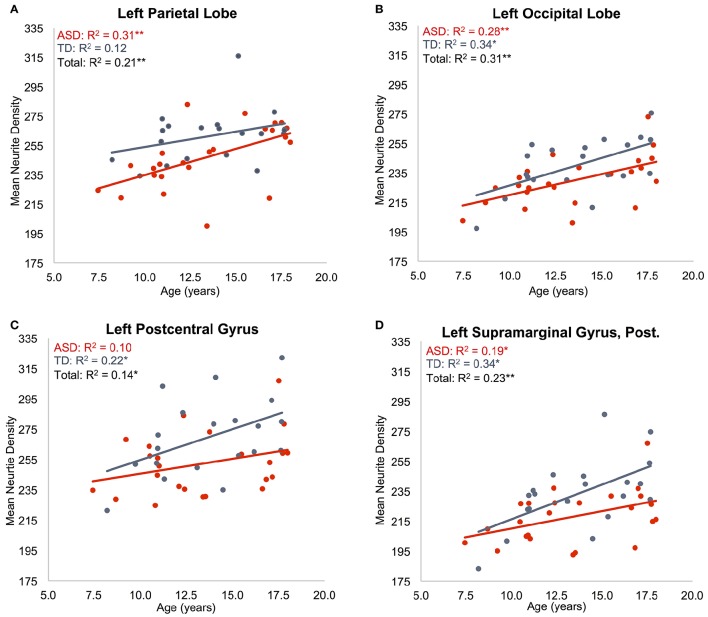
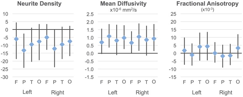
Autism postmortem studies have shown various cytoarchitectural anomalies in cortical and limbic areas including increased cell packing density, laminar disorganization, and narrowed minicolumns. However, there is little evidence on dendritic and axonal organization in ASD. Recent imaging techniques have the potential for non-invasive, studies of small-scale structure in the human brain, including gray matter. Here, Restriction Spectrum Imaging (RSI), a multi-shell diffusion-weighted imaging technique, was used to examine gray matter microstructure in 24 children with ASD (5 female) and 20 matched typically developing (TD) participants (2 female), ages 7-17 years. RSI extends the spherical deconvolution model to multiple length scales to characterize neurite density (ND) and organization. Measures were examined in 48 cortical regions of interest per hemisphere. To our knowledge, this is the first time that a multi-compartmental diffusion model has been applied to cortical gray matter in ASD. The ND measure detected robust age effects showing a significant positive relationship to age in all lobes except left temporal when groups were combined. Results were also suggestive of group differences (ASD<TD) in anterior cingulate, right superior temporal lobe and much of the parietal lobes, but these fell short of statistical significance. For MD, significant group differences (ASD>TD) in bilateral parietal regions as well as widespread age effects were detected. Our findings support the value of multi-shell diffusion imaging for assays of cortical gray matter. This approach has the potential to add to postmortem literature, examining intracortical organization, intracortical axonal content, myelination, or caliber. Robust age effects further support the validity of the ND metric for examination of gray matter microstructure in ASD and across development. While diffusion MRI does not approach the precision of histological studies, imaging measures of microstructure can complement postmortem studies, by allowing access to large sample sizes, a whole-brain field of view, longitudinal designs, and combination with behavioral and functional assays. This makes multi-shell diffusion imaging a promising technique for understanding the underlying cytoarchitecture of the disorder.
自闭症的尸检研究表明,皮质和边缘区域存在各种细胞结构异常,包括细胞堆积密度增加、层状结构紊乱和微小柱变窄。然而,关于自闭症谱系障碍(ASD)中树突和轴突组织的证据很少。最近的成像技术有潜力对包括灰质在内的人脑小尺度结构进行非侵入性研究。在这里,限制谱成像(RSI),一种多壳扩散加权成像技术,被用于检查24名患有ASD的儿童(5名女性)和20名年龄匹配的发育正常(TD)参与者(2名女性)的灰质微观结构,年龄在7至17岁之间。RSI将球形去卷积模型扩展到多个长度尺度,以表征神经突密度(ND)和组织。在每个半球的48个皮质感兴趣区域进行测量。据我们所知,这是首次将多室扩散模型应用于ASD患者的皮质灰质。ND测量检测到显著的年龄效应,表明在合并组时,除左颞叶外,所有脑叶中ND与年龄呈显著正相关。结果还提示在前扣带回、右颞上叶和大部分顶叶存在组间差异(ASD<TD),但这些差异未达到统计学显著性。对于平均扩散率(MD),在双侧顶叶区域检测到显著的组间差异(ASD>TD)以及广泛的年龄效应。我们的研究结果支持多壳扩散成像在皮质灰质检测中的价值。这种方法有可能补充尸检文献,用于检查皮质内组织、皮质内轴突含量、髓鞘形成或管径。显著的年龄效应进一步支持了ND指标在检查ASD患者及整个发育过程中灰质微观结构方面的有效性。虽然扩散磁共振成像(MRI)无法达到组织学研究的精度,但微观结构的成像测量可以通过允许获取大样本量、全脑视野、纵向设计以及与行为和功能检测相结合,来补充尸检研究。这使得多壳扩散成像成为理解该疾病潜在细胞结构的一种有前途的技术。