• 文献检索
  • 文档翻译
  • 深度研究
  • 学术资讯
  • Suppr Zotero 插件Zotero 插件
  • 邀请有礼
  • 套餐&价格
  • 历史记录
应用&插件
Suppr Zotero 插件Zotero 插件浏览器插件Mac 客户端Windows 客户端微信小程序
定价
高级版会员购买积分包购买API积分包
服务
文献检索文档翻译深度研究API 文档MCP 服务
关于我们
关于 Suppr公司介绍联系我们用户协议隐私条款
关注我们

Suppr 超能文献

核心技术专利:CN118964589B侵权必究
粤ICP备2023148730 号-1Suppr @ 2026

文献检索

告别复杂PubMed语法,用中文像聊天一样搜索,搜遍4000万医学文献。AI智能推荐,让科研检索更轻松。

立即免费搜索

文件翻译

保留排版,准确专业,支持PDF/Word/PPT等文件格式,支持 12+语言互译。

免费翻译文档

深度研究

AI帮你快速写综述,25分钟生成高质量综述,智能提取关键信息,辅助科研写作。

立即免费体验

全脑磁共振成像图谱:对现有图谱及在人群成像中使用的注意事项的系统综述。

Whole Brain Magnetic Resonance Image Atlases: A Systematic Review of Existing Atlases and Caveats for Use in Population Imaging.

作者信息

Dickie David Alexander, Shenkin Susan D, Anblagan Devasuda, Lee Juyoung, Blesa Cabez Manuel, Rodriguez David, Boardman James P, Waldman Adam, Job Dominic E, Wardlaw Joanna M

机构信息

Brain Research Imaging Centre, Neuroimaging Sciences, Centre for Clinical Brain Sciences, Royal Infirmary of Edinburgh, The University of EdinburghEdinburgh, UK; Scottish Imaging Network, A Platform for Scientific Excellence (SINAPSE) CollaborationGlasgow, UK.

Brain Research Imaging Centre, Neuroimaging Sciences, Centre for Clinical Brain Sciences, Royal Infirmary of Edinburgh, The University of EdinburghEdinburgh, UK; Geriatric Medicine Unit, Royal Infirmary of Edinburgh, The University of EdinburghEdinburgh, UK; Department of Psychology, Centre for Cognitive Ageing and Cognitive Epidemiology, The University of EdinburghEdinburgh, UK.

出版信息

Front Neuroinform. 2017 Jan 19;11:1. doi: 10.3389/fninf.2017.00001. eCollection 2017.

DOI:10.3389/fninf.2017.00001
PMID:28154532
原文链接:https://pmc.ncbi.nlm.nih.gov/articles/PMC5244468/
Abstract

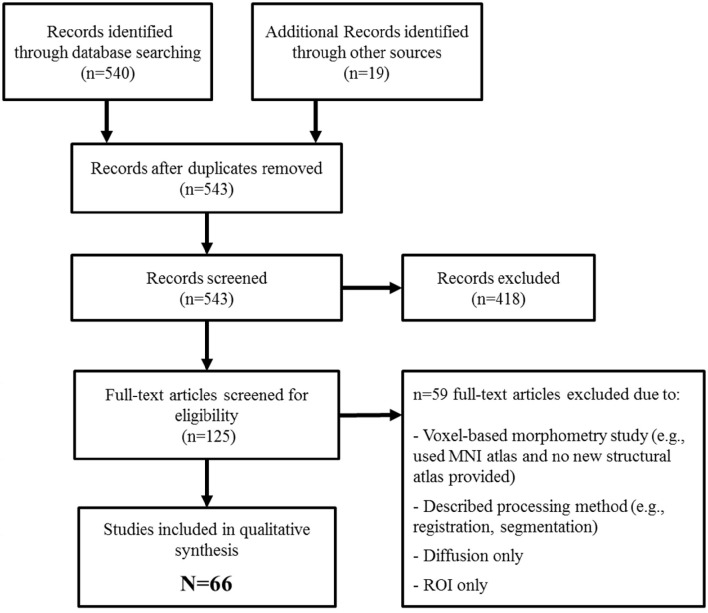
Brain MRI atlases may be used to characterize brain structural changes across the life course. Atlases have important applications in research, e.g., as registration and segmentation targets to underpin image analysis in population imaging studies, and potentially in future in clinical practice, e.g., as templates for identifying brain structural changes out with normal limits, and increasingly for use in surgical planning. However, there are several caveats and limitations which must be considered before successfully applying brain MRI atlases to research and clinical problems. For example, the influential Talairach and Tournoux atlas was derived from a single fixed cadaveric brain from an elderly female with limited clinical information, yet is the basis of many modern atlases and is often used to report locations of functional activation. We systematically review currently available whole brain structural MRI atlases with particular reference to the implications for population imaging through to emerging clinical practice. We found 66 whole brain structural MRI atlases world-wide. The vast majority were based on T1, T2, and/or proton density (PD) structural sequences, had been derived using parametric statistics (inappropriate for brain volume distributions), had limited supporting clinical or cognitive data, and included few younger (>5 and <18 years) or older (>60 years) subjects. To successfully characterize brain structural features and their changes across different stages of life, we conclude that whole brain structural MRI atlases should include: more subjects at the upper and lower extremes of age; additional structural sequences, including fluid attenuation inversion recovery (FLAIR) and T2 sequences; a range of appropriate statistics, e.g., rank-based or non-parametric; and detailed cognitive and clinical profiles of the included subjects in order to increase the relevance and utility of these atlases.

摘要

脑磁共振成像图谱可用于描述一生中脑结构的变化。图谱在研究中具有重要应用,例如作为配准和分割目标,以支持人群成像研究中的图像分析,并且在未来可能应用于临床实践,例如作为识别超出正常范围的脑结构变化的模板,以及越来越多地用于手术规划。然而,在成功将脑磁共振成像图谱应用于研究和临床问题之前,必须考虑几个注意事项和局限性。例如,有影响力的Talairach和Tournoux图谱源自一名老年女性的单一固定尸体脑,临床信息有限,但却是许多现代图谱的基础,并且经常用于报告功能激活的位置。我们系统地综述了目前可用的全脑结构磁共振成像图谱,特别提及了对从人群成像到新兴临床实践的影响。我们发现全球有66种全脑结构磁共振成像图谱。绝大多数基于T1、T2和/或质子密度(PD)结构序列,使用参数统计方法得出(不适用于脑体积分布),支持的临床或认知数据有限,并且包括的年轻(>5岁和<18岁)或年长(>60岁)受试者较少。为了成功描述不同生命阶段的脑结构特征及其变化,我们得出结论,全脑结构磁共振成像图谱应包括:更多年龄上限和下限的受试者;额外的结构序列,包括液体衰减反转恢复(FLAIR)和T2序列;一系列适当的统计方法,例如基于秩或非参数的方法;以及所纳入受试者的详细认知和临床概况,以提高这些图谱的相关性和实用性。

https://cdn.ncbi.nlm.nih.gov/pmc/blobs/d288/5244468/45a06390c707/fninf-11-00001-g0001.jpg

相似文献

1
Whole Brain Magnetic Resonance Image Atlases: A Systematic Review of Existing Atlases and Caveats for Use in Population Imaging.全脑磁共振成像图谱:对现有图谱及在人群成像中使用的注意事项的系统综述。
Front Neuroinform. 2017 Jan 19;11:1. doi: 10.3389/fninf.2017.00001. eCollection 2017.
2
Multi-atlas segmentation of the whole hippocampus and subfields using multiple automatically generated templates.使用多个自动生成的模板对整个海马体及其子区进行多图谱分割。
Neuroimage. 2014 Nov 1;101:494-512. doi: 10.1016/j.neuroimage.2014.04.054. Epub 2014 Apr 29.
3
Use of brain MRI atlases to determine boundaries of age-related pathology: the importance of statistical method.利用脑磁共振成像图谱确定年龄相关病理学的边界:统计方法的重要性。
PLoS One. 2015 May 29;10(5):e0127939. doi: 10.1371/journal.pone.0127939. eCollection 2015.
4
BRAHMA: Population specific T1, T2, and FLAIR weighted brain templates and their impact in structural and functional imaging studies.Brahma:特定人群的 T1、T2 和 FLAIR 加权脑模板及其对结构和功能成像研究的影响。
Magn Reson Imaging. 2020 Jul;70:5-21. doi: 10.1016/j.mri.2019.12.009. Epub 2020 Jan 7.
5
Structural brain atlases: design, rationale, and applications in normal and pathological cohorts.结构脑图谱:设计、原理及在正常和病理队列中的应用。
J Alzheimers Dis. 2012;31 Suppl 3(0 3):S169-88. doi: 10.3233/JAD-2012-120412.
6
Non-parametric MRI Brain Atlas for the Polish Population.波兰人群的非参数MRI脑图谱。
Front Neuroinform. 2021 Oct 6;15:684759. doi: 10.3389/fninf.2021.684759. eCollection 2021.
7
Unbiased age-specific structural brain atlases for Chinese pediatric population.针对中国儿童群体的无偏结构脑图谱
Neuroimage. 2019 Apr 1;189:55-70. doi: 10.1016/j.neuroimage.2019.01.006. Epub 2019 Jan 6.
8
Construction of multi-region-multi-reference atlases for neonatal brain MRI segmentation.构建新生儿脑 MRI 分割的多区域-多参考图谱。
Neuroimage. 2010 Jun;51(2):684-93. doi: 10.1016/j.neuroimage.2010.02.025. Epub 2010 Feb 17.
9
A multi-atlas based method for automated anatomical Macaca fascicularis brain MRI segmentation and PET kinetic extraction.基于多图谱的方法用于自动解剖猕猴大脑 MRI 分割和 PET 动力学提取。
Neuroimage. 2013 Aug 15;77:26-43. doi: 10.1016/j.neuroimage.2013.03.029. Epub 2013 Mar 26.
10
Stereotaxic Magnetic Resonance Imaging Brain Atlases for Infants from 3 to 12 Months.3至12个月婴儿的立体定向磁共振成像脑图谱
Dev Neurosci. 2015;37(6):515-32. doi: 10.1159/000438749. Epub 2015 Oct 7.

引用本文的文献

1
Longitudinal diffusion tensor imaging correlates with amyloid burden in Down syndrome.纵向扩散张量成像与唐氏综合征中的淀粉样蛋白负荷相关。
Alzheimers Dement. 2025 Aug;21(8):e70572. doi: 10.1002/alz.70572.
2
Deep-Diffeomorphic Networks for Conditional Brain Templates.用于条件脑模板的深度微分同胚网络
Hum Brain Mapp. 2025 Jun 1;46(8):e70229. doi: 10.1002/hbm.70229.
3
Quantifying Arm and Leg Movements in 3-Month-Old Infants Using Pose Estimation: Proof of Concept.使用姿势估计对3个月大婴儿的手臂和腿部运动进行量化:概念验证。

本文引用的文献

1
Human Connectomics across the Life Span.人类全生命周期连接组学
Trends Cogn Sci. 2017 Jan;21(1):32-45. doi: 10.1016/j.tics.2016.10.005. Epub 2016 Nov 16.
2
Multimodal population brain imaging in the UK Biobank prospective epidemiological study.英国生物银行前瞻性流行病学研究中的多模态人群脑成像
Nat Neurosci. 2016 Nov;19(11):1523-1536. doi: 10.1038/nn.4393. Epub 2016 Sep 19.
3
A multi-modal parcellation of human cerebral cortex.人类大脑皮层的多模态分区
Sensors (Basel). 2024 Nov 27;24(23):7586. doi: 10.3390/s24237586.
4
Super-resolution multi-contrast unbiased eye atlases with deep probabilistic refinement.具有深度概率细化的超分辨率多对比度无偏眼图谱
J Med Imaging (Bellingham). 2024 Nov;11(6):064004. doi: 10.1117/1.JMI.11.6.064004. Epub 2024 Nov 14.
5
Predicting high-level visual areas in the absence of task fMRI.在没有任务 fMRI 的情况下预测高级视觉区域。
Sci Rep. 2024 May 18;14(1):11376. doi: 10.1038/s41598-024-62098-9.
6
Fetal brain MRI atlases and datasets: A review.胎儿脑 MRI 图谱和数据集:综述。
Neuroimage. 2024 Apr 15;292:120603. doi: 10.1016/j.neuroimage.2024.120603. Epub 2024 Apr 6.
7
Impact of brain parcellation on prediction performance in models of cognition and demographics.脑区划分对认知和人口统计学模型预测性能的影响。
Hum Brain Mapp. 2024 Feb 1;45(2):e26592. doi: 10.1002/hbm.26592.
8
Anatomical registration of intracranial electrodes. Robust model-based localization and deformable smooth brain-shift compensation methods.颅内电极的解剖配准。基于稳健模型的定位和可变形平滑脑移位补偿方法。
J Neurosci Methods. 2024 Apr;404:110056. doi: 10.1016/j.jneumeth.2024.110056. Epub 2024 Jan 14.
9
Joint cortical registration of geometry and function using semi-supervised learning.使用半监督学习的几何与功能联合皮质配准
ArXiv. 2023 Oct 16:arXiv:2303.01592v4.
10
A personalized cortical atlas for functional regions of interest.用于功能感兴趣区的个性化皮质图谱。
J Neurophysiol. 2023 Nov 1;130(5):1067-1080. doi: 10.1152/jn.00108.2023. Epub 2023 Sep 20.
Nature. 2016 Aug 11;536(7615):171-178. doi: 10.1038/nature18933. Epub 2016 Jul 20.
4
Parcellation of the Healthy Neonatal Brain into 107 Regions Using Atlas Propagation through Intermediate Time Points in Childhood.通过儿童期中间时间点的图谱传播将健康新生儿脑部分割为107个区域
Front Neurosci. 2016 May 19;10:220. doi: 10.3389/fnins.2016.00220. eCollection 2016.
5
The Human Brainnetome Atlas: A New Brain Atlas Based on Connectional Architecture.人类脑网络组图谱:基于连接结构的新脑图谱。
Cereb Cortex. 2016 Aug;26(8):3508-26. doi: 10.1093/cercor/bhw157. Epub 2016 May 26.
6
Vascular risk factors and progression of white matter hyperintensities in the Lothian Birth Cohort 1936.1936年洛锡安出生队列中的血管危险因素与白质高信号的进展
Neurobiol Aging. 2016 Jun;42:116-23. doi: 10.1016/j.neurobiolaging.2016.03.011. Epub 2016 Mar 21.
7
A brain imaging repository of normal structural MRI across the life course: Brain Images of Normal Subjects (BRAINS).一个涵盖整个生命历程的正常结构磁共振成像的脑成像数据库:正常受试者脑图像(BRAINS)。
Neuroimage. 2017 Jan;144(Pt B):299-304. doi: 10.1016/j.neuroimage.2016.01.027. Epub 2016 Jan 18.
8
Brain volumetric changes and cognitive ageing during the eighth decade of life.八十岁年龄段的脑容量变化与认知衰老
Hum Brain Mapp. 2015 Dec;36(12):4910-25. doi: 10.1002/hbm.22959. Epub 2015 Sep 7.
9
Progression of White Matter Disease and Cortical Thinning Are Not Related in Older Community-Dwelling Subjects.在社区居住的老年人中,白质病变进展与皮质变薄无关。
Stroke. 2016 Feb;47(2):410-6. doi: 10.1161/STROKEAHA.115.011229. Epub 2015 Dec 22.
10
Resource atlases for multi-atlas brain segmentations with multiple ontology levels based on T1-weighted MRI.基于T1加权磁共振成像的具有多个本体层次的多图谱脑部分割资源图谱集。
Neuroimage. 2016 Jan 15;125:120-130. doi: 10.1016/j.neuroimage.2015.10.042. Epub 2015 Oct 21.