Glasser Matthew F, Coalson Timothy S, Robinson Emma C, Hacker Carl D, Harwell John, Yacoub Essa, Ugurbil Kamil, Andersson Jesper, Beckmann Christian F, Jenkinson Mark, Smith Stephen M, Van Essen David C
Department of Neuroscience, Washington University Medical School, Saint Louis, Missouri 63110, USA.
FMRIB centre, Nuffield Department of Clinical Neurosciences, John Radcliffe Hospital, University of Oxford, Oxford OX3 9DU, UK.
Nature. 2016 Aug 11;536(7615):171-178. doi: 10.1038/nature18933. Epub 2016 Jul 20.
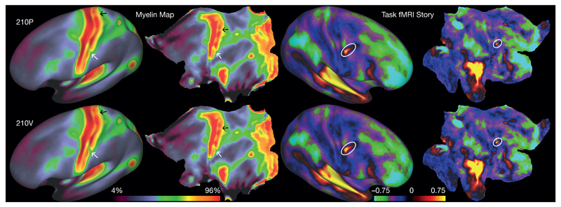
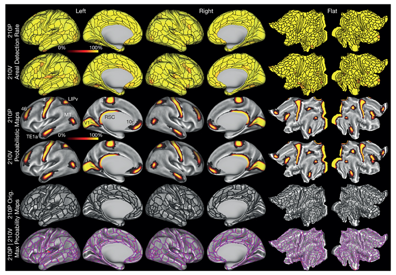
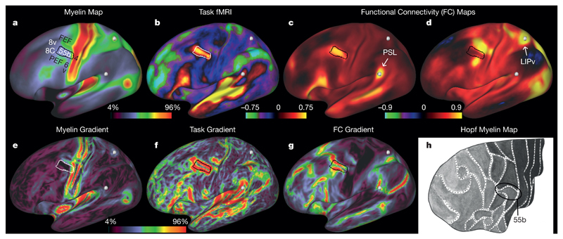
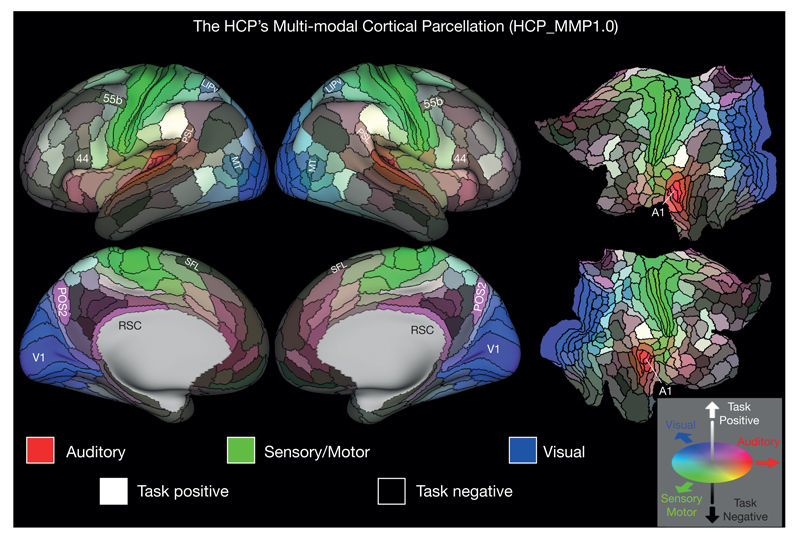
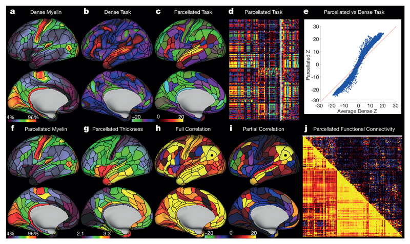
Understanding the amazingly complex human cerebral cortex requires a map (or parcellation) of its major subdivisions, known as cortical areas. Making an accurate areal map has been a century-old objective in neuroscience. Using multi-modal magnetic resonance images from the Human Connectome Project (HCP) and an objective semi-automated neuroanatomical approach, we delineated 180 areas per hemisphere bounded by sharp changes in cortical architecture, function, connectivity, and/or topography in a precisely aligned group average of 210 healthy young adults. We characterized 97 new areas and 83 areas previously reported using post-mortem microscopy or other specialized study-specific approaches. To enable automated delineation and identification of these areas in new HCP subjects and in future studies, we trained a machine-learning classifier to recognize the multi-modal 'fingerprint' of each cortical area. This classifier detected the presence of 96.6% of the cortical areas in new subjects, replicated the group parcellation, and could correctly locate areas in individuals with atypical parcellations. The freely available parcellation and classifier will enable substantially improved neuroanatomical precision for studies of the structural and functional organization of human cerebral cortex and its variation across individuals and in development, aging, and disease.
要理解极其复杂的人类大脑皮层,需要绘制其主要亚区(即皮质区域)的图谱(或进行分区)。绘制精确的区域图谱一直是神经科学领域一个世纪以来的目标。利用来自人类连接组计划(HCP)的多模态磁共振图像以及一种客观的半自动神经解剖学方法,我们在210名健康年轻成年人精确对齐的群体平均值中,勾勒出每个半球180个由皮质结构、功能、连接性和/或地形的急剧变化所界定的区域。我们对97个新区域以及之前使用尸检显微镜或其他特定研究专用方法报道过的83个区域进行了特征描述。为了能够在新的HCP受试者以及未来的研究中自动勾勒和识别这些区域,我们训练了一个机器学习分类器来识别每个皮质区域的多模态“指纹”。该分类器在新受试者中检测到了96.6%的皮质区域,复制了群体分区,并且能够在具有非典型分区的个体中正确定位区域。免费提供的分区和分类器将极大提高神经解剖学精度,用于研究人类大脑皮层的结构和功能组织及其在个体间以及发育、衰老和疾病过程中的变化。