Sun Jun, Yan Songhua, Jiang Yan, Wong Duo Wai-Chi, Zhang Ming, Zeng Jizhou, Zhang Kuan
School of Biomedical Engineering, Capital Medical University, Beijing, 100069, China.
Beijing Key Laboratory of Fundamental Research on Biomechanics in Clinical Application, Capital Medical University, Beijing, 100069, China.
Biomed Eng Online. 2016 Dec 28;15(Suppl 2):158. doi: 10.1186/s12938-016-0253-3.
Knee valgus and varus morbidity is at the second top place in children lower limb deformity diseases. It may cause abnormal stress distribution. The magnitude and location of contact forces on tibia plateau during gait cycle have been indicated as markers for risk of osteoarthritis. So far, few studies reported the contact stress and force distribution on tibial plateau of valgus knee of children.
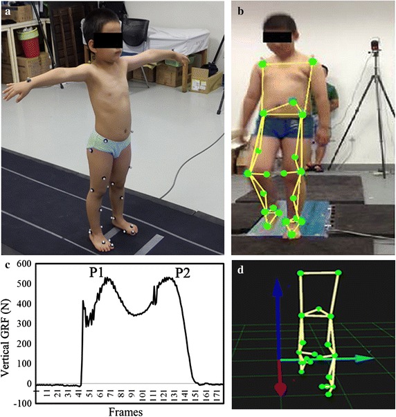
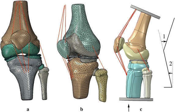
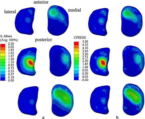
To estimate the contact stresses and forces on tibial plateau of an 8-year old obese boy with valgus knee and a 7-year old healthy boy, three-dimensional (3D) finite element (FE) models of their left knee joints were developed. The valgus knee model has 36,897 nodes and 1,65,106 elements, and the normal knee model has 78,278 nodes and 1,18,756 elements. Paired t test was used for the comparison between the results from the 3D FE analysis method and the results from traditional kinematic measurement methods.
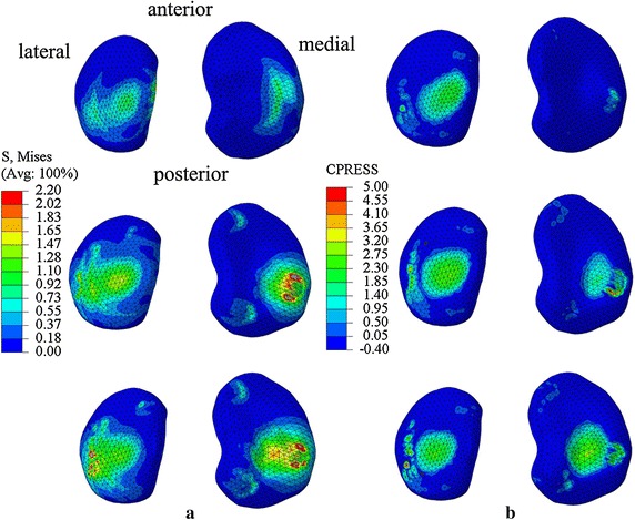
The p value of paired t test is 0.12. Maximum stresses shifted to lateral plateau in knee valgus children while maximum stresses were on medial plateau in normal knee child at the first peak of vertical GRF of stance phase. The locations of contact centers on medial plateau changed 3.38 mm more than that on lateral plateau, while the locations of contact centers on medial plateau changed 1.22 mm less than that on lateral plateau for healthy child from the first peak to second peak of vertical GRF of stance phase.
The paired t test result shows that there is no significant difference between the two methods. The results of FE analysis method suggest that knee valgus malalignment could be the reason for abnormal knee load that may cause knee problems in obese children with valgus knee in the long-term. This study may help to understand biomechanical mechanism of valgus knees of obese children.
膝外翻和膝内翻发病率在儿童下肢畸形疾病中位居第二。它可能导致异常的应力分布。步态周期中胫骨平台上接触力的大小和位置已被视为骨关节炎风险的标志物。到目前为止,很少有研究报道儿童膝外翻时胫骨平台的接触应力和力分布情况。
为了评估一名8岁肥胖膝外翻男孩和一名7岁健康男孩胫骨平台上的接触应力和力,建立了他们左膝关节的三维(3D)有限元(FE)模型。膝外翻模型有36897个节点和165106个单元,正常膝关节模型有78278个节点和118756个单元。采用配对t检验对三维有限元分析方法的结果与传统运动学测量方法的结果进行比较。
配对t检验的p值为0.12。在站立相垂直地面反力的第一个峰值时,膝外翻儿童的最大应力转移到外侧平台,而正常膝关节儿童的最大应力在内侧平台。在站立相垂直地面反力从第一个峰值到第二个峰值的过程中,膝外翻儿童内侧平台接触中心的位置变化比外侧平台多3.38毫米,而健康儿童内侧平台接触中心的位置变化比外侧平台少1.22毫米。
配对t检验结果表明两种方法之间无显著差异。有限元分析方法的结果表明,膝外翻畸形可能是导致肥胖膝外翻儿童膝关节负荷异常的原因,长期来看可能会引发膝关节问题。本研究可能有助于理解肥胖儿童膝外翻的生物力学机制。