Kieffer Collin, Ladinsky Mark S, Ninh Allen, Galimidi Rachel P, Bjorkman Pamela J
Division of Biology and Biological Engineering, California Institute of Technology, Pasadena, United States.
Elife. 2017 Feb 15;6:e23282. doi: 10.7554/eLife.23282.
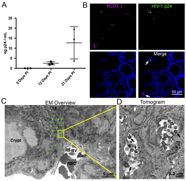
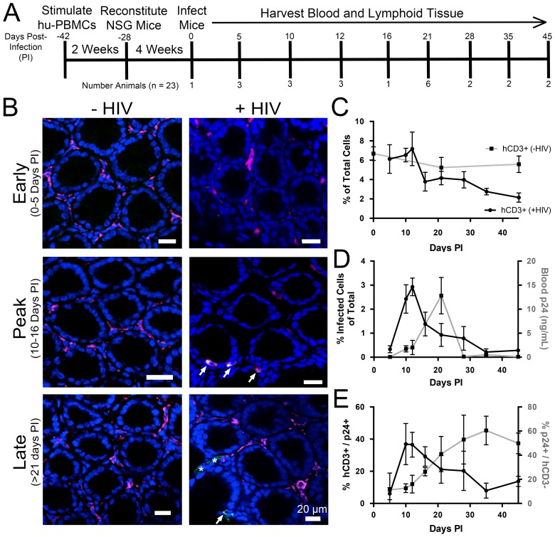
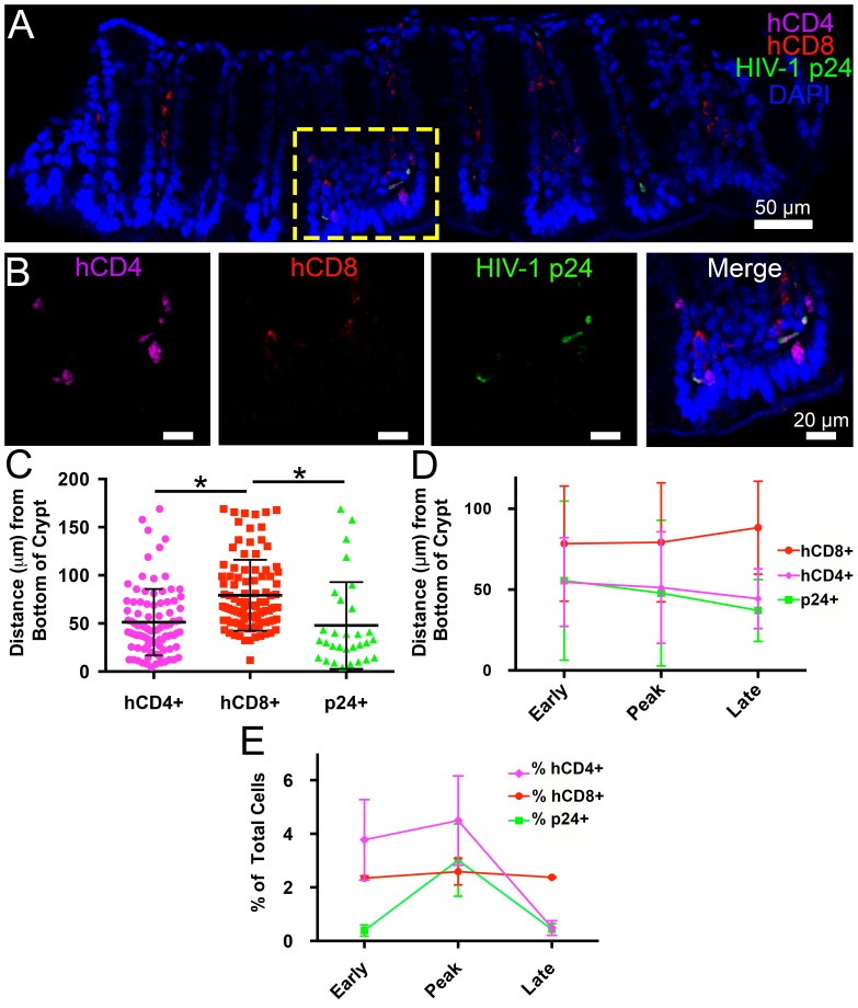
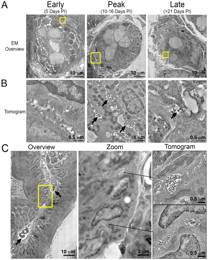
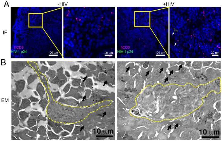
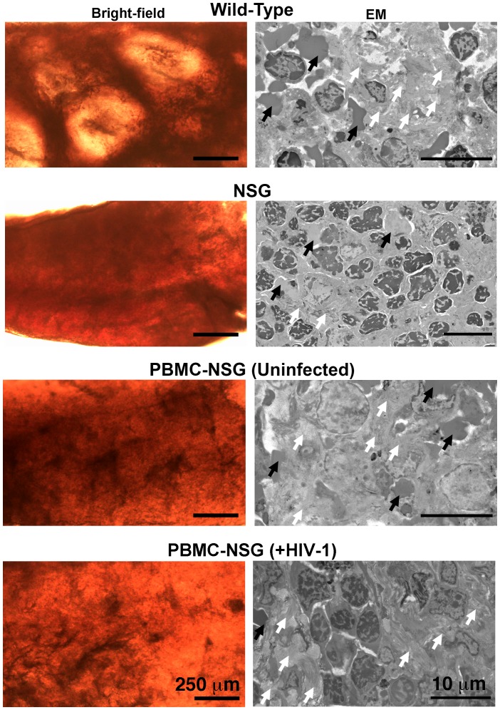
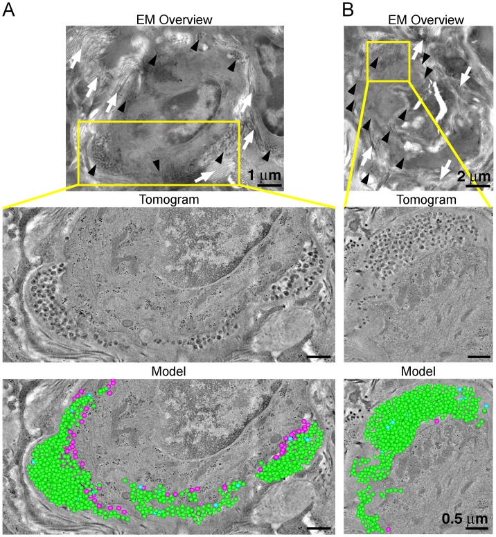
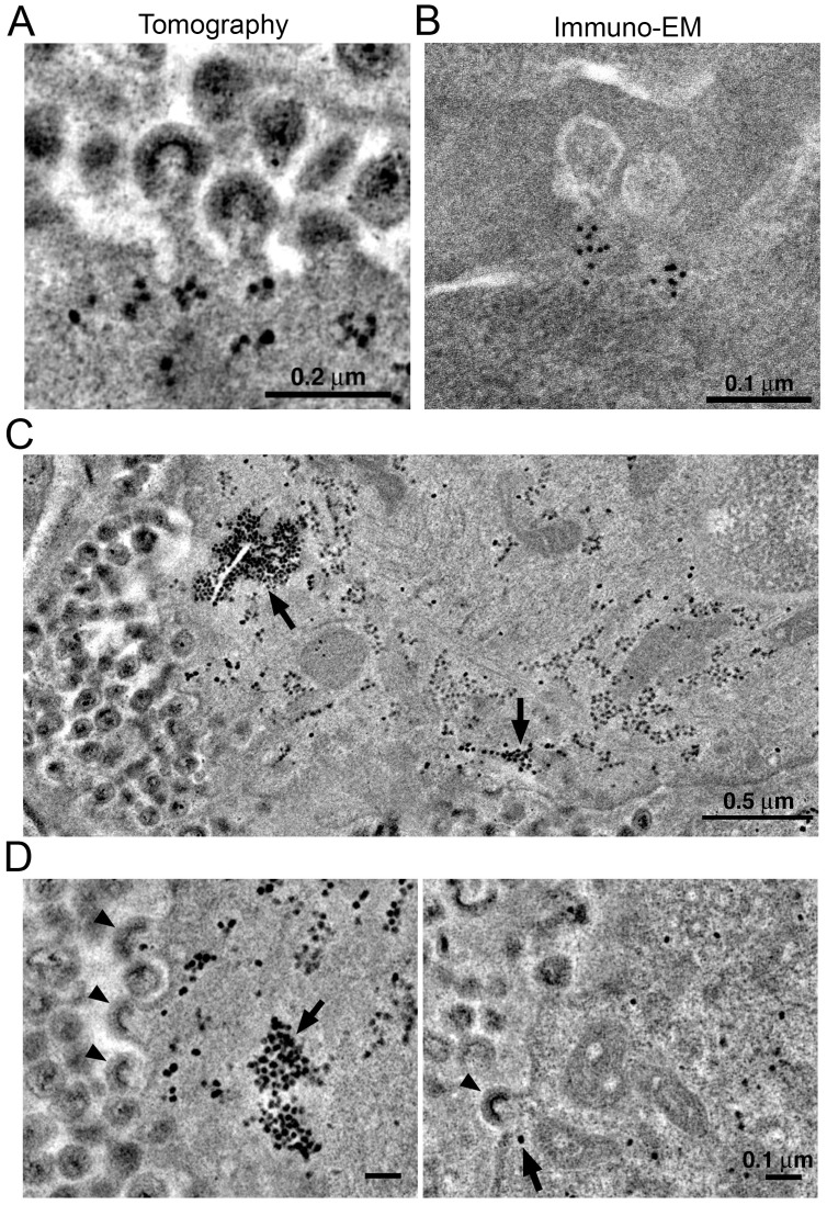
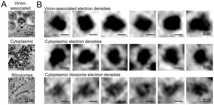
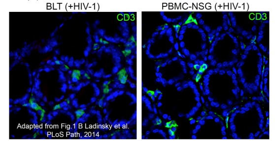
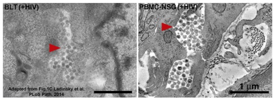
Dissemination of HIV-1 throughout lymphoid tissues leads to systemic virus spread following infection. We combined tissue clearing, 3D-immunofluorescence, and electron tomography (ET) to longitudinally assess early HIV-1 spread in lymphoid tissues in humanized mice. Immunofluorescence revealed peak infection density in gut at 10-12 days post-infection when blood viral loads were low. Human CD4+ T-cells and HIV-1-infected cells localized predominantly to crypts and the lower third of intestinal villi. Free virions and infected cells were not readily detectable by ET at 5-days post-infection, whereas HIV-1-infected cells surrounded by pools of free virions were present in ~10% of intestinal crypts by 10-12 days. ET of spleen revealed thousands of virions released by individual cells and discreet cytoplasmic densities near sites of prolific virus production. These studies highlight the importance of multiscale imaging of HIV-1-infected tissues and are adaptable to other animal models and human patient samples.
HIV-1在整个淋巴组织中的传播会导致感染后病毒在全身扩散。我们结合组织透明化、三维免疫荧光和电子断层扫描(ET),纵向评估人源化小鼠淋巴组织中HIV-1的早期传播情况。免疫荧光显示,在感染后10-12天,当血液病毒载量较低时,肠道中的感染密度达到峰值。人CD4+ T细胞和HIV-1感染细胞主要定位于隐窝和肠绒毛的下三分之一处。在感染后5天,通过ET不易检测到游离病毒粒子和感染细胞,而到10-12天时,约10%的肠隐窝中存在被游离病毒粒子池包围的HIV-1感染细胞。脾脏的ET显示,单个细胞释放出数千个病毒粒子,并且在大量产生病毒的部位附近存在离散的细胞质密度。这些研究突出了对HIV-1感染组织进行多尺度成像的重要性,并且适用于其他动物模型和人类患者样本。