Department of Chemical Biology & Drug Discovery, Utrecht Institute for Pharmaceutical Sciences, Utrecht University, Utrecht, The Netherlands.
Department of Translational Neuroscience, University Medical Center Utrecht Brain Center, Utrecht University, Utrecht, The Netherlands.
PLoS Pathog. 2022 Mar 7;18(3):e1010340. doi: 10.1371/journal.ppat.1010340. eCollection 2022 Mar.
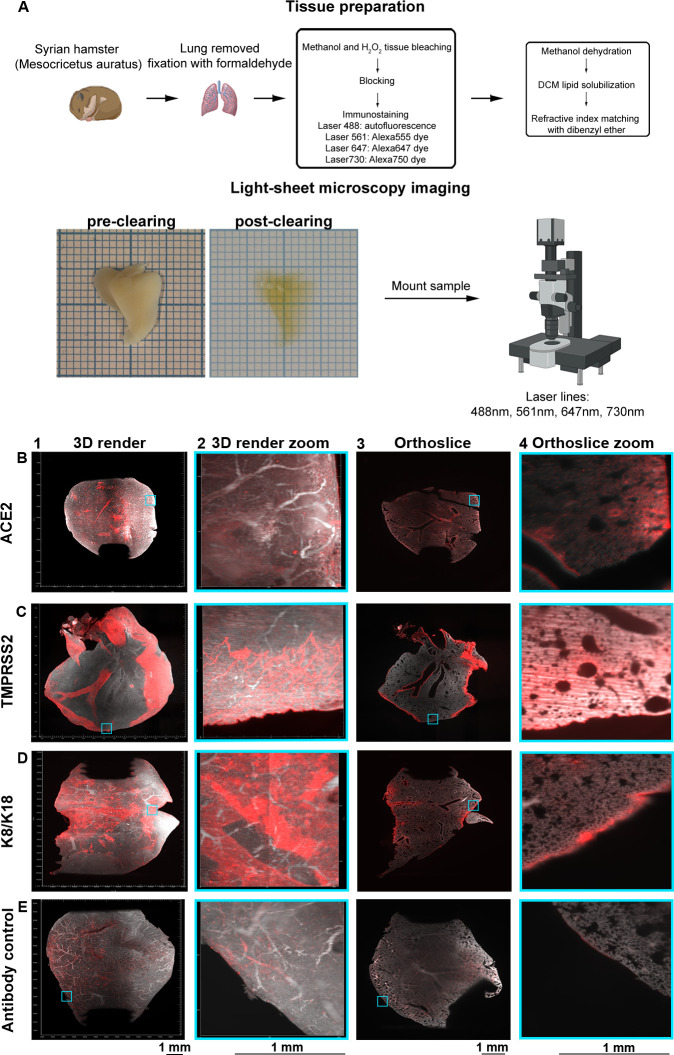
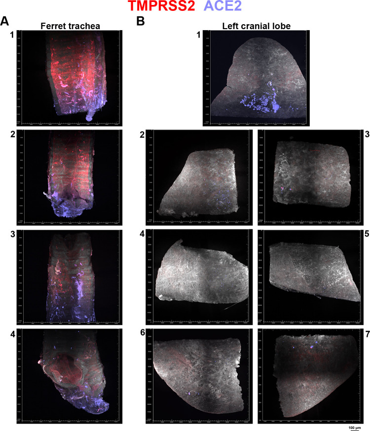
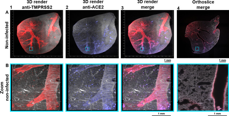
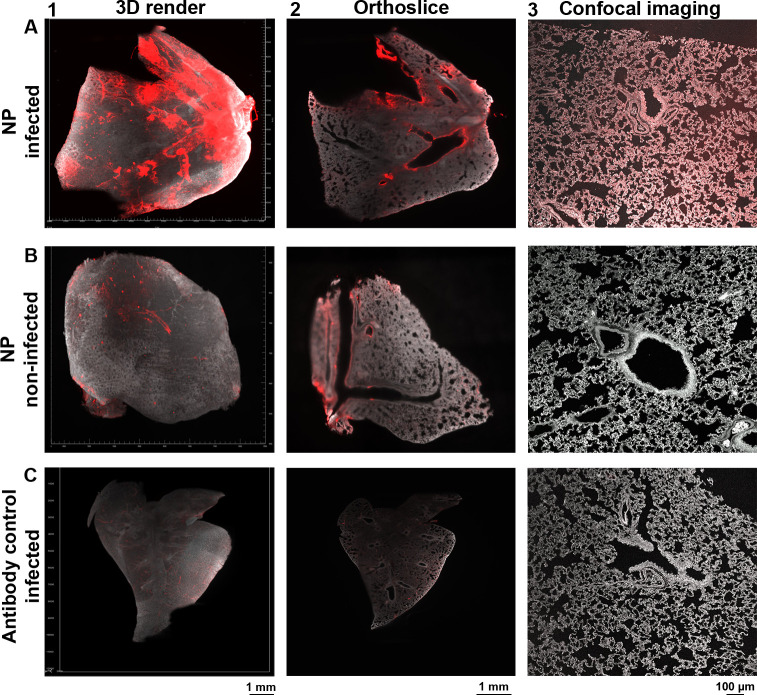
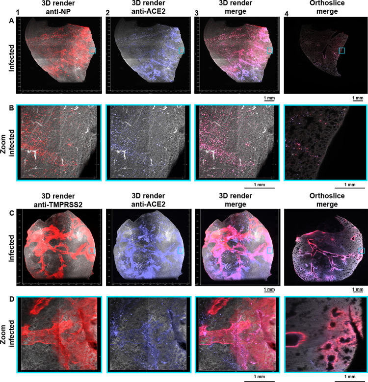
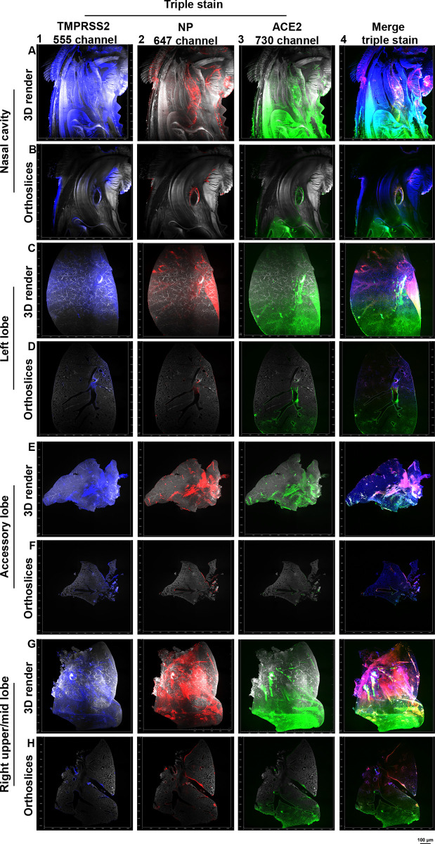
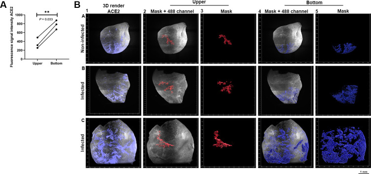
SARS-CoV-2 attaches to angiotensin-converting enzyme 2 (ACE2) to gain entry into cells after which the spike protein is cleaved by the transmembrane serine protease 2 (TMPRSS2) to facilitate viral-host membrane fusion. ACE2 and TMPRSS2 expression profiles have been analyzed at the genomic, transcriptomic, and single-cell RNAseq levels. However, transcriptomic data and actual protein validation convey conflicting information regarding the distribution of the biologically relevant protein receptor in whole tissues. To describe the organ-level architecture of receptor expression, related to the ability of ACE2 and TMPRSS2 to mediate infectivity, we performed a volumetric analysis of whole Syrian hamster lung lobes. Lung tissue of infected and control animals was stained using antibodies against ACE2 and TMPRSS2, combined with SARS-CoV-2 nucleoprotein staining. This was followed by light-sheet microscopy imaging to visualize their expression and related infection patterns. The data demonstrate that infection is restricted to sites containing both ACE2 and TMPRSS2, the latter is expressed in the primary and secondary bronchi whereas ACE2 is predominantly observed in the bronchioles and alveoli. Conversely, infection completely overlaps where ACE2 and TMPRSS2 co-localize in the tertiary bronchi, bronchioles, and alveoli.
SARS-CoV-2 通过血管紧张素转化酶 2(ACE2)进入细胞,随后通过跨膜丝氨酸蛋白酶 2(TMPRSS2)切割刺突蛋白,促进病毒-宿主膜融合。已经在基因组、转录组和单细胞 RNAseq 水平分析了 ACE2 和 TMPRSS2 的表达谱。然而,转录组数据和实际蛋白质验证传达了关于整个组织中生物相关蛋白受体分布的相互矛盾的信息。为了描述与 ACE2 和 TMPRSS2 介导感染性相关的受体表达的器官水平结构,我们对感染和对照动物的整个叙利亚仓鼠肺叶进行了容积分析。使用针对 ACE2 和 TMPRSS2 的抗体对肺组织进行染色,结合 SARS-CoV-2 核蛋白染色。随后进行光片显微镜成像以可视化它们的表达和相关的感染模式。数据表明,感染仅限于同时包含 ACE2 和 TMPRSS2 的部位,后者在一级和二级支气管中表达,而 ACE2 主要在细支气管和肺泡中观察到。相反,感染完全重叠 ACE2 和 TMPRSS2 在三级支气管、细支气管和肺泡中共定位的部位。