Lebedeva Aleksandra K, Westman Eric, Borza Tom, Beyer Mona K, Engedal Knut, Aarsland Dag, Selbaek Geir, Haberg Asta K
Department of Neurobiology, Care Sciences and Society, Karolinska Institutet Stockholm, Sweden.
Centre for Old Age Psychiatric Research, Innlandet Hospital TrustBrumunddal, Norway; Institute of Clinical Medicine, Faculty of Medicine, University of OsloOslo, Norway.
Front Aging Neurosci. 2017 Feb 2;9:13. doi: 10.3389/fnagi.2017.00013. eCollection 2017.
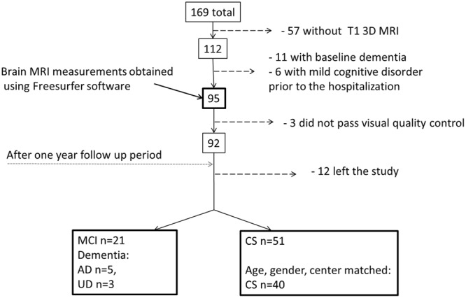
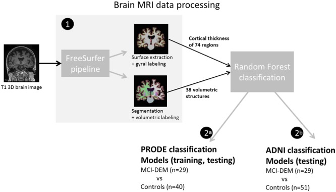
Late-life depression (LLD) is associated with development of different types of dementia. Identification of LLD patients, who will develop cognitive decline, i.e., the early stage of dementia would help to implement interventions earlier. The purpose of this study was to assess whether structural brain magnetic resonance imaging (MRI) in LLD patients can predict mild cognitive impairment (MCI) or dementia 1 year prior to the diagnosis. LLD patients underwent brain MRI at baseline and repeated clinical assessment after 1-year. Structural brain measurements were obtained using Freesurfer software (v. 5.1) from the T1W brain MRI images. MRI-based Random Forest classifier was used to discriminate between LLD who developed MCI or dementia after 1-year follow-up and cognitively stable LLD. Additionally, a previously established Random Forest model trained on 185 patients with Alzheimer's disease (AD) vs. 225 cognitively normal elderly from the Alzheimer's disease Neuroimaging Initiative was tested on the LLD data set (ADNI model). MCI and dementia diagnoses were predicted in LLD patients with 76%/68%/84% accuracy/sensitivity/specificity. Adding the baseline Mini-Mental State Examination (MMSE) scores to the models improved accuracy/sensitivity/specificity to 81%/75%/86%. The best model predicted MCI status alone using MRI and baseline MMSE scores with accuracy/sensitivity/specificity of 89%/85%/90%. The most important region for all the models was right ventral diencephalon, including hypothalamus. Its volume correlated negatively with the number of depressive episodes. ADNI model trained on AD vs. Controls using SV could predict MCI-DEM patients with 67% accuracy. LDD patients developing MCI and dementia can be discriminated from LLD patients remaining cognitively stable with good accuracy based on baseline structural MRI alone. Baseline MMSE score improves prediction accuracy. Ventral diencephalon, including the hypothalamus might play an important role in preservation of cognitive functions in LLD.
老年期抑郁症(LLD)与不同类型痴呆症的发生有关。识别出将会出现认知衰退(即痴呆症早期)的LLD患者,有助于更早地实施干预措施。本研究的目的是评估LLD患者的脑部结构磁共振成像(MRI)是否能够在诊断前1年预测轻度认知障碍(MCI)或痴呆症。LLD患者在基线时接受脑部MRI检查,并在1年后进行重复临床评估。使用Freesurfer软件(版本5.1)从T1加权脑部MRI图像中获取脑部结构测量数据。基于MRI的随机森林分类器用于区分在1年随访后发展为MCI或痴呆症的LLD患者和认知稳定的LLD患者。此外,在一组来自阿尔茨海默病神经影像倡议组织的185例阿尔茨海默病(AD)患者与225例认知正常老年人身上训练的一个先前建立的随机森林模型,在LLD数据集上进行了测试(ADNI模型)。在LLD患者中预测MCI和痴呆症诊断的准确率/灵敏度/特异度分别为76%/68%/84%。将基线简易精神状态检查表(MMSE)评分添加到模型中后,准确率/灵敏度/特异度提高到了81%/75%/86%。最佳模型仅使用MRI和基线MMSE评分预测MCI状态,准确率/灵敏度/特异度为89%/85%/90%。所有模型中最重要的区域是右腹侧间脑,包括下丘脑。其体积与抑郁发作次数呈负相关。使用支持向量机在AD与对照人群上训练的ADNI模型预测MCI - DEM患者的准确率为67%。仅基于基线结构MRI,就能够以较高的准确率区分发展为MCI和痴呆症的LLD患者与认知保持稳定的LLD患者。基线MMSE评分可提高预测准确率。包括下丘脑在内的腹侧间脑可能在LLD患者认知功能的维持中发挥重要作用。