Ilie Marius, Szafer-Glusman Edith, Hofman Véronique, Long-Mira Elodie, Suttmann Rebecca, Darbonne Walter, Butori Catherine, Lalvée Salomé, Fayada Julien, Selva Eric, Yu Wei, Marquette Charles-Hugo, Shames David S, Punnoose Elizabeth, Hofman Paul
Laboratory of Clinical and Experimental Pathology and Liquid Biopsy Laboratory, Pasteur Hospital, University Hospital Federation OncoAge, Université Côte d'Azur, Nice, France.
Institute for Research on Cancer and Ageing, Nice (IRCAN), INSERM U1081/UMR CNRS 7284, Team 3, Antoine Lacassagne Cancer Center, Nice, France.
Oncotarget. 2017 Apr 18;8(16):26112-26121. doi: 10.18632/oncotarget.15345.
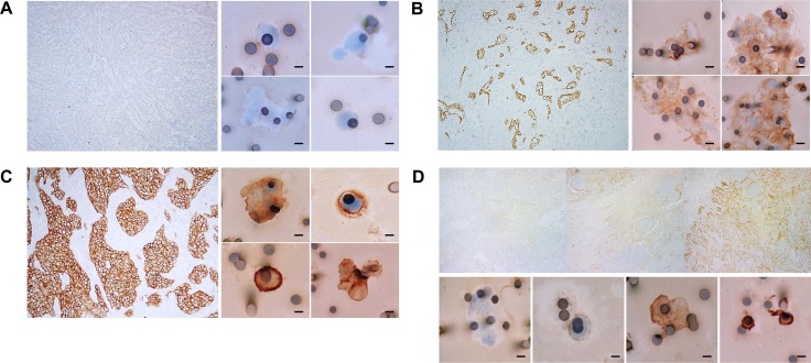
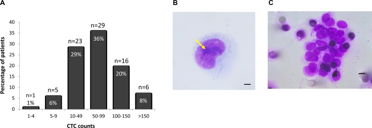
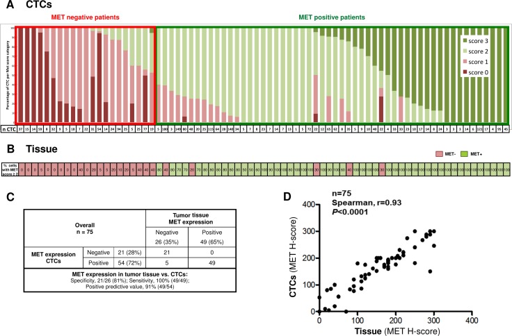
Given the difficulty in obtaining adequate tissue in NSCLC, we investigated the utility of circulating tumor cells (CTCs) for MET status assessment in NSCLC patients. We used two platforms for CTC capture, and assessed MET expression in CTCs and matched-bronchial biopsies in patients with advanced-stage III/IV lung adenocarcinoma. Baseline peripheral blood was collected from 256 advanced-stage III/IV NSCLC patients from Genentech clinical trials, and from 106 patients with advanced-stage III/IV lung adenocarcinoma treated at the Department of Pneumology, Pasteur Hospital, Nice. CTCs were enriched using CellSearch (Genentech), or ISET technologies (Pasteur Hospital). MET expression was evaluated by immunofluorescence on CellSearch, and by immunocytochemistry on ISET-enriched CTCs and on matched FFPE tissue sections (Pasteur Hospital). CTCs were detected in 83 of 256 (32%) patients evaluated on CellSearch, with 30 samples (12%) exhibiting ≥ 5 CTCs/7.5 ml blood. On ISET, CTC were observed in 80 of 106 patients (75%), and 79 patients (75%) exhibited ≥ 5 CTCs/4 ml blood. MET expression on ISET CTCs was positive in 72% of cases, and the MET expression on matched-patient tissue was positive in 65% patients using the Onartuzumab IHC scoring algorithm (93% concordance). Quantification of MET expression using H-scores showed strong correlation between MET expression in tissue and CTCs (Spearman correlation, 0.93). MET status in CTCs isolated on ISET filters from blood samples of advanced-stage NSCLC patients correlated strongly with MET status in tumor tissue, illustrating the potential for using CTCs as a non-invasive, real-time biopsy to determine MET status of patients entering clinical trials.
鉴于在非小细胞肺癌(NSCLC)中获取足够组织存在困难,我们研究了循环肿瘤细胞(CTC)在NSCLC患者MET状态评估中的效用。我们使用了两种平台来捕获CTC,并评估了晚期III/IV期肺腺癌患者CTC中的MET表达以及匹配的支气管活检组织中的MET表达。从基因泰克公司临床试验的256例晚期III/IV期NSCLC患者以及尼斯巴斯德医院肺病科治疗的106例晚期III/IV期肺腺癌患者中采集基线外周血。使用CellSearch(基因泰克公司)或ISET技术(巴斯德医院)富集CTC。通过CellSearch上的免疫荧光以及ISET富集的CTC和匹配的福尔马林固定石蜡包埋(FFPE)组织切片(巴斯德医院)上的免疫细胞化学评估MET表达。在使用CellSearch评估的256例患者中的83例(32%)检测到CTC,其中30个样本(12%)每7.5毫升血液中显示≥5个CTC。在ISET上,106例患者中的80例(75%)观察到CTC,79例患者(75%)每4毫升血液中显示≥5个CTC。使用Onartuzumab免疫组化评分算法,ISET CTC上的MET表达在72%的病例中呈阳性,匹配患者组织上的MET表达在65%的患者中呈阳性(一致性为93%)。使用H评分对MET表达进行定量显示组织和CTC中的MET表达之间存在强相关性(Spearman相关性,0.93)。从晚期NSCLC患者血液样本的ISET滤器上分离的CTC中的MET状态与肿瘤组织中的MET状态密切相关,这表明使用CTC作为非侵入性实时活检来确定进入临床试验患者的MET状态具有潜力。