Gustafsson Johan, Sundlöv Anna, Sjögreen Gleisner Katarina
Department of Medical Radiation Physics, Clinical Sciences Lund, Lund University, Lund, Sweden.
Department of Oncology and Pathology, Clinical Sciences Lund, Lund University, Lund, Sweden.
EJNMMI Res. 2017 Dec;7(1):18. doi: 10.1186/s13550-017-0262-7. Epub 2017 Feb 23.
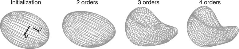
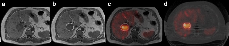
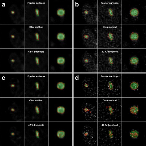
Dosimetry in radionuclide therapy has the potential to allow for a treatment tailored to the individual patient. One therapeutic radiopharmaceutical where patient-specific dosimetry is feasible is Lu-DOTATATE, used for the treatment of neuroendocrine tumours. The emission of gamma photons by Lu allows for imaging with SPECT (single photon emission computed tomography). One important step for dosimetry using this imaging technique is the SPECT image segmentation, which needs to be robust and accurate for the estimated quantities to be reliable. This work investigates different methods for automatic tumour delineation in Lu-DOTATATE SPECT images. Three segmentation methods are considered: a fixed 42% threshold (FT), the Otsu method (OM) and a method based on Fourier surfaces (FS). Effects of including resolution compensation in the iterative SPECT image reconstruction are also studied. Evaluation is performed based on Monte Carlo-simulated SPECT images from 24 h and 336 h post injection (p.i.), for determination of the volume, activity concentration and dice similarity coefficient. In addition, patient data are used to investigate the correspondence of tumour volumes when delineated in SPECT or morphological CT or MR images. Patient data are also used to examine the sensitivity to the operator-dependent initialization.
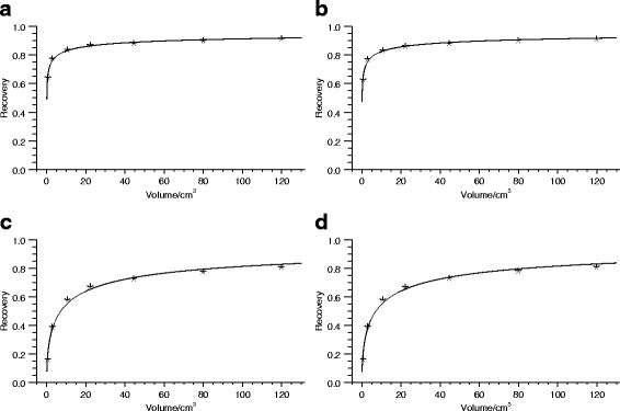
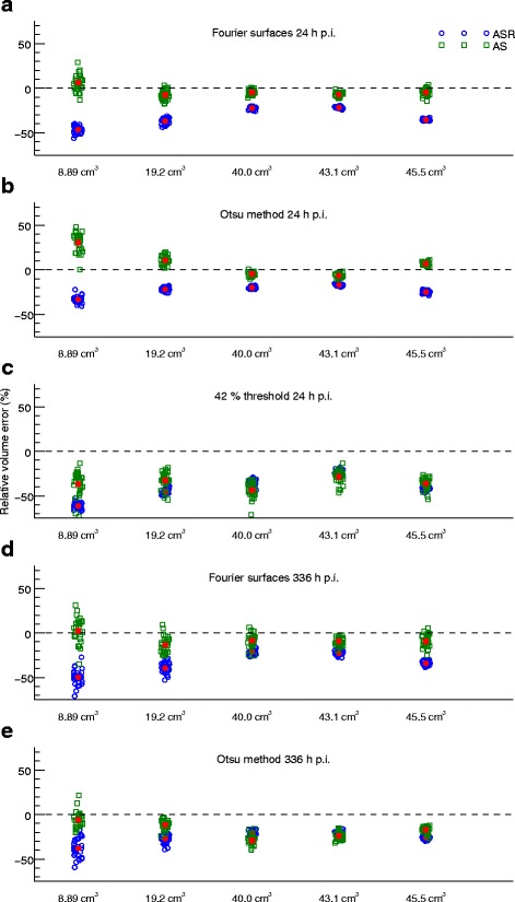
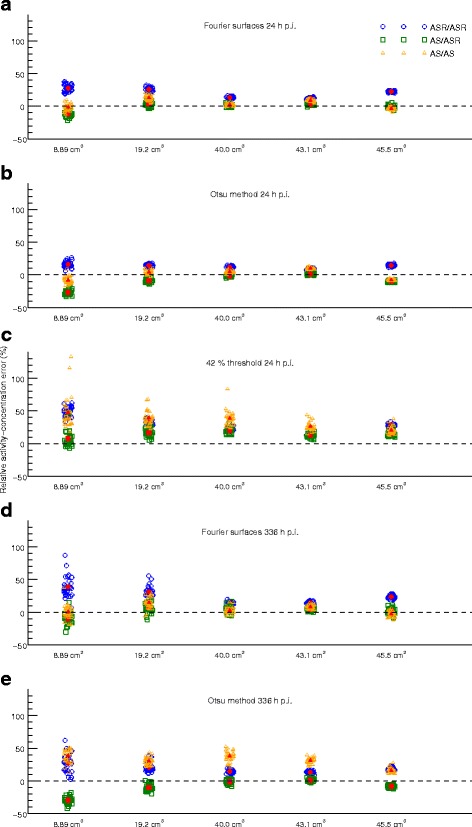
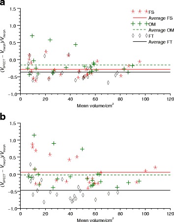
For simulated images from 24 h p.i. reconstructed without resolution compensation, a volume and activity-concentration root-mean-square error below 15% is typically obtained for tumours above approximately 10 cm when using OM or FS, while FT performs considerably worse. When including resolution compensation, the tumour volume becomes underestimated and the activity concentration overestimated. The FS method appears to be robust to noise, as seen for the 336 h images. The differences between the tumour volumes estimated from the SPECT images and the volumes estimated from morphological images are generally larger than the discrepancies seen for the simulated data sets.
Segmentation results are encouraging for future dosimetry of tumours with volumes above approximately 10 cm. Using resolution compensation in the reconstruction may have a negative effect on volume estimation.
放射性核素治疗中的剂量测定有潜力实现针对个体患者的个性化治疗。一种可行的针对患者的剂量测定的治疗性放射性药物是用于治疗神经内分泌肿瘤的镥 - DOTATATE。镥发射的伽马光子可用于单光子发射计算机断层扫描(SPECT)成像。使用这种成像技术进行剂量测定的一个重要步骤是SPECT图像分割,分割需要稳健且准确,以便估计的量可靠。这项工作研究了镥 - DOTATATE SPECT图像中自动肿瘤勾勒的不同方法。考虑了三种分割方法:固定的42%阈值(FT)、大津法(OM)和基于傅里叶曲面的方法(FS)。还研究了在迭代SPECT图像重建中纳入分辨率补偿的效果。基于注射后(p.i.)24小时和336小时的蒙特卡罗模拟SPECT图像进行评估,以确定体积、活度浓度和骰子相似系数。此外,使用患者数据来研究在SPECT、形态学CT或MR图像中勾勒出的肿瘤体积的对应关系。患者数据还用于检查对操作者依赖的初始化的敏感性。
对于注射后24小时重建且未进行分辨率补偿的模拟图像,当使用OM或FS时,对于大约10 cm以上的肿瘤,通常可获得低于15%的体积和活度浓度均方根误差,而FT的表现则差得多。当纳入分辨率补偿时,肿瘤体积被低估,活度浓度被高估。如336小时图像所示,FS方法似乎对噪声具有鲁棒性。从SPECT图像估计的肿瘤体积与从形态学图像估计的体积之间的差异通常大于模拟数据集的差异。
对于体积约10 cm以上的肿瘤,未来剂量测定的分割结果令人鼓舞。在重建中使用分辨率补偿可能对体积估计产生负面影响。