Yang Chunguang, Ma Xueyou, Wang Zhihua, Zeng Xing, Hu Zhiquan, Ye Zhangqun, Shen Guanxin
Department of Urology, Tongji Hospital.
Department of Immunology, Tongji Medical College, Huazhong University of Science and Technology, Wuhan, Hubei, People's Republic of China.
Drug Des Devel Ther. 2017 Feb 14;11:431-439. doi: 10.2147/DDDT.S126964. eCollection 2017.
Curcumin induces apoptosis and autophagy in different cancer cells. Moreover, chemical and biological experiments have evidenced that curcumin is a biologically active iron chelator and induces cytotoxicity through iron chelation. We thus hypothesized that curcumin may induce apoptosis and autophagy in castration-resistant prostate cancer (CRPC) cells through its iron-chelating properties.
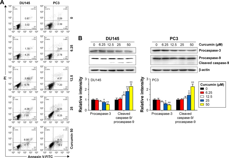
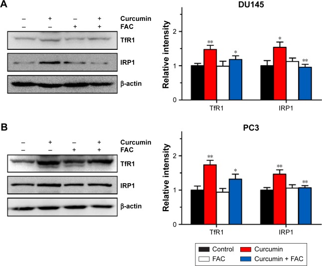
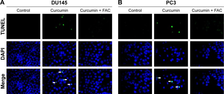
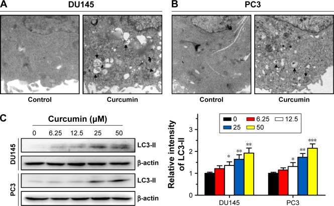
CRPC cells were loaded with curcumin alone or in combination with ferric ammonium citrate (FAC). Cytotoxicity was measured by 3-(4,5-dimethylthiazol-2-yl)-2,5-diphenyltetrazolium bromide (MTT) assay. Apoptosis was assessed by flow cytometry, terminal deoxynucleotidyl transferase nick end labeling (TUNEL) assay and caspase activity. Autophagy status was analyzed by the detection of autophagosomes and light chain 3-II (LC3-II) using transmission electron microscopy and Western blot. Iron-binding activity of curcumin was assessed by spectrophotometry and MTT assay. The expression levels of transferrin receptor 1 (TfR1) and iron regulatory protein 1 (IRP1) were examined by Western blot.
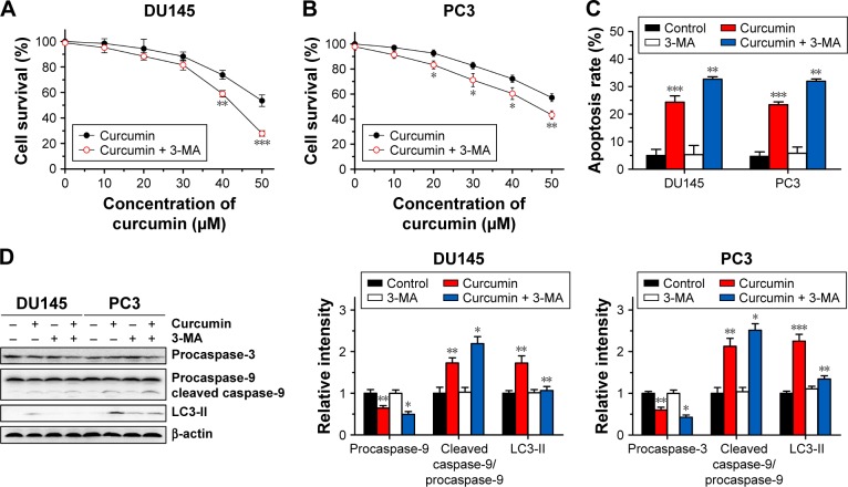
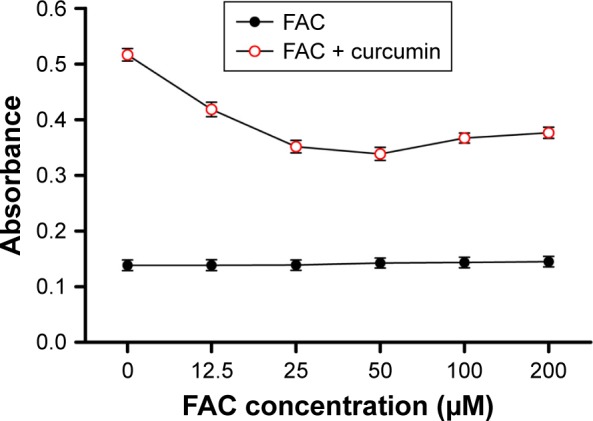
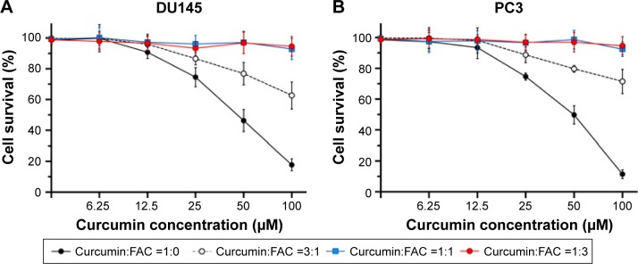
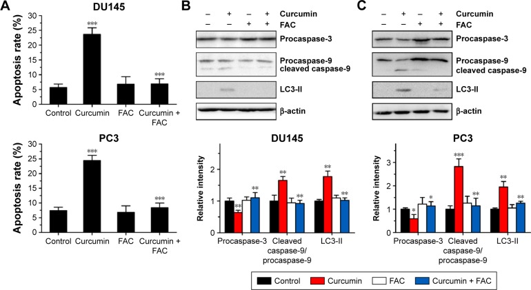
Curcumin induced apoptosis and autophagy in CRPC cells. Combining curcumin with autophagy inhibitors (3-methyladenine [3-MA]) synergized the apoptotic effect of curcumin. Moreover, curcumin bound to FAC at a ratio of ~1:1, as assessed by spectrophotometry and MTT assay. Apoptosis and autophagy induced by curcumin were counteracted by equal amounts of FAC. At apoptosis- and autophagy-inducing concentrations, curcumin enhanced the expression levels of TfR1 and IRP1, indicative of iron deprivation induced by curcumin.
Together, our results indicate that curcumin induces apoptosis and protective autophagy in CRPC cells, which are at least partially dependent on its iron-chelating properties.
姜黄素可诱导不同癌细胞发生凋亡和自噬。此外,化学和生物学实验已证明姜黄素是一种具有生物活性的铁螯合剂,并通过铁螯合诱导细胞毒性。因此,我们推测姜黄素可能通过其铁螯合特性诱导去势抵抗性前列腺癌(CRPC)细胞发生凋亡和自噬。
CRPC细胞单独或与柠檬酸铁铵(FAC)联合加载姜黄素。通过3-(4,5-二甲基噻唑-2-基)-2,5-二苯基四氮唑溴盐(MTT)法检测细胞毒性。通过流式细胞术、末端脱氧核苷酸转移酶介导的缺口末端标记(TUNEL)法和半胱天冬酶活性评估细胞凋亡。使用透射电子显微镜和蛋白质免疫印迹法通过检测自噬体和轻链3-II(LC3-II)分析自噬状态。通过分光光度法和MTT法评估姜黄素的铁结合活性。通过蛋白质免疫印迹法检测转铁蛋白受体1(TfR1)和铁调节蛋白1(IRP1)的表达水平。
姜黄素诱导CRPC细胞发生凋亡和自噬。将姜黄素与自噬抑制剂(3-甲基腺嘌呤[3-MA])联合使用可增强姜黄素的凋亡作用。此外,通过分光光度法和MTT法评估,姜黄素与FAC的结合比例约为1:1。等量的FAC可抵消姜黄素诱导的凋亡和自噬。在诱导凋亡和自噬的浓度下,姜黄素提高了TfR1和IRP1的表达水平,表明姜黄素诱导了铁缺乏。
总之,我们的结果表明姜黄素在CRPC细胞中诱导凋亡和保护性自噬,这至少部分依赖于其铁螯合特性。