Zhou Zhi-Peng, Long Li-Ling, Qiu Wei-Jia, Cheng Ge, Huang Li-Juan, Yang Teng-Fei, Huang Zhong-Kui
Department of Radiology, The First Affiliated Hospital of Guangxi Medical University, Nanning, Guangxi, 530021, People's Republic of China.
Department of Radiology, Affiliated Hospital of Guilin Medical University, Guilin, Guangxi, 541001, People's Republic of China.
BMC Med Imaging. 2017 Mar 1;17(1):20. doi: 10.1186/s12880-017-0192-x.
Assessing the liver function provides valuable information to evaluate surgical risk and plan accordingly. Current studies focus on whole liver function evaluation. However, assessment of segmental liver function is equally important in the clinical practice. The purpose of this study was to investigate whether Gd-EOB-DTPA-enhanced MRI can evaluate the liver function of each segment by using T1 mapping at 3 Tesla MRI.
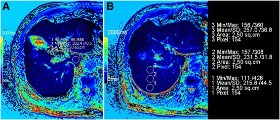
One hundred three patients were classified into one of 4 groups: a normal liver function (NLF) group (n = 38), a liver cirrhosis with Child-Pugh A (LCA) group (n = 33), a liver cirrhosis with Child-Pugh B (LCB) group (n = 21), and a liver cirrhosis with Child-Pugh C (LCC) group (n = 11). All patients underwent Gd-EOB-DTPA-enhanced MRI scans. T1 relaxation times were measured on the liver superimposing T1 mapping images. Reduction rate (△%) of T1 relaxation time of the liver parenchyma were calculated.
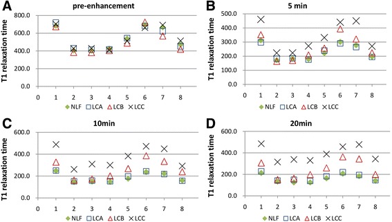
After 20 min of Gd-EOB-DTPA enhancement, the T1 relaxation time of all liver segments in the LCC group were different from those in all the other groups, and more liver segments from the LCB and LCA groups different from the NLF group (p < 0.05). For the LCB group, the areas under the receiver operating characteristic curves (AUCs) of different liver segments for hepatobiliary phase (HBP) were 0.654-0.904 on T1 relaxation time, and 0.709-0.905 on △%. For the LCC group, the AUCs of different liver segments for HBP were 0.842-0.997 on T1 relaxation time, and 0.887-0.990 on △%.
For LCB patients, segmental liver function evaluation is possible using Gd-EOB-DTPA-enhanced MRI T1 mapping. For LCC patients, all liver segments can be used to evaluate liver function and both T1 relaxation time and the △% of T1 relaxation time have good diagnostic performance.
评估肝功能可为评估手术风险并据此制定计划提供有价值的信息。目前的研究集中在全肝功能评估上。然而,在临床实践中,节段性肝功能评估同样重要。本研究的目的是探讨钆塞酸二钠增强磁共振成像(Gd-EOB-DTPA-enhanced MRI)能否在3特斯拉磁共振成像中通过T1映射评估各节段的肝功能。
103例患者被分为4组之一:正常肝功能(NLF)组(n = 38)、Child-Pugh A级肝硬化(LCA)组(n = 33)、Child-Pugh B级肝硬化(LCB)组(n = 21)和Child-Pugh C级肝硬化(LCC)组(n = 11)。所有患者均接受钆塞酸二钠增强磁共振成像扫描。在叠加T1映射图像的肝脏上测量T1弛豫时间。计算肝实质T1弛豫时间的降低率(△%)。
钆塞酸二钠增强20分钟后,LCC组所有肝段的T1弛豫时间与其他所有组不同,LCB和LCA组更多肝段与NLF组不同(p < 0.05)。对于LCB组,肝胆期(HBP)不同肝段在T1弛豫时间上的受试者操作特征曲线下面积(AUC)为0.654 - 0.904,在△%上为0.709 - 0.905。对于LCC组,HBP不同肝段在T1弛豫时间上的AUC为0.842 - 0.997,在△%上为0.887 - 0.990。
对于LCB患者,使用钆塞酸二钠增强磁共振成像T1映射可以进行节段性肝功能评估。对于LCC患者,所有肝段均可用于评估肝功能,T1弛豫时间和T1弛豫时间降低率(△%)均具有良好的诊断性能。