Lee Dong-Kyu, Ahn Kyung-Sik, Kang Chang Ho, Cho Sung Bum
Department of Radiology, Korea University Anam Hospital, Korea University College of Medicine, Seoul, Korea.
Ultrasonography. 2017 Apr;36(2):120-130. doi: 10.14366/usg.17001. Epub 2017 Jan 30.
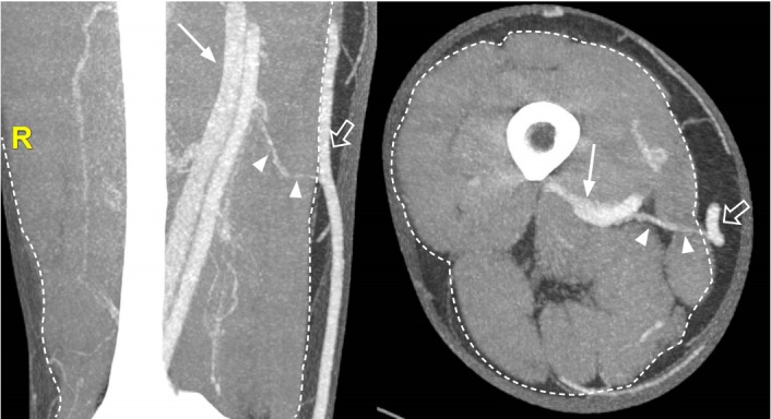
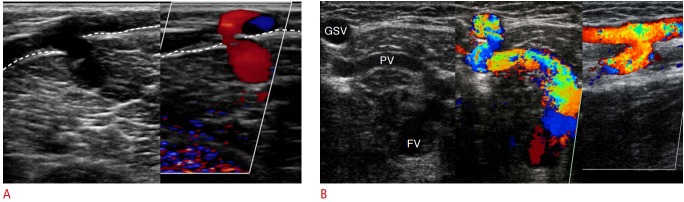
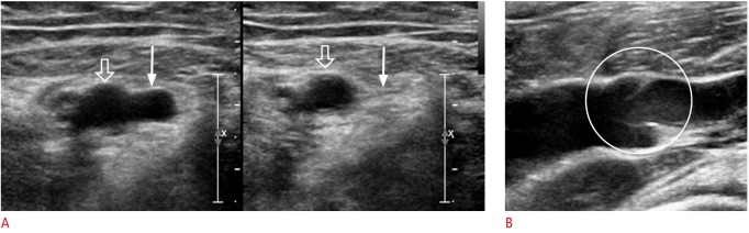
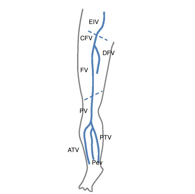
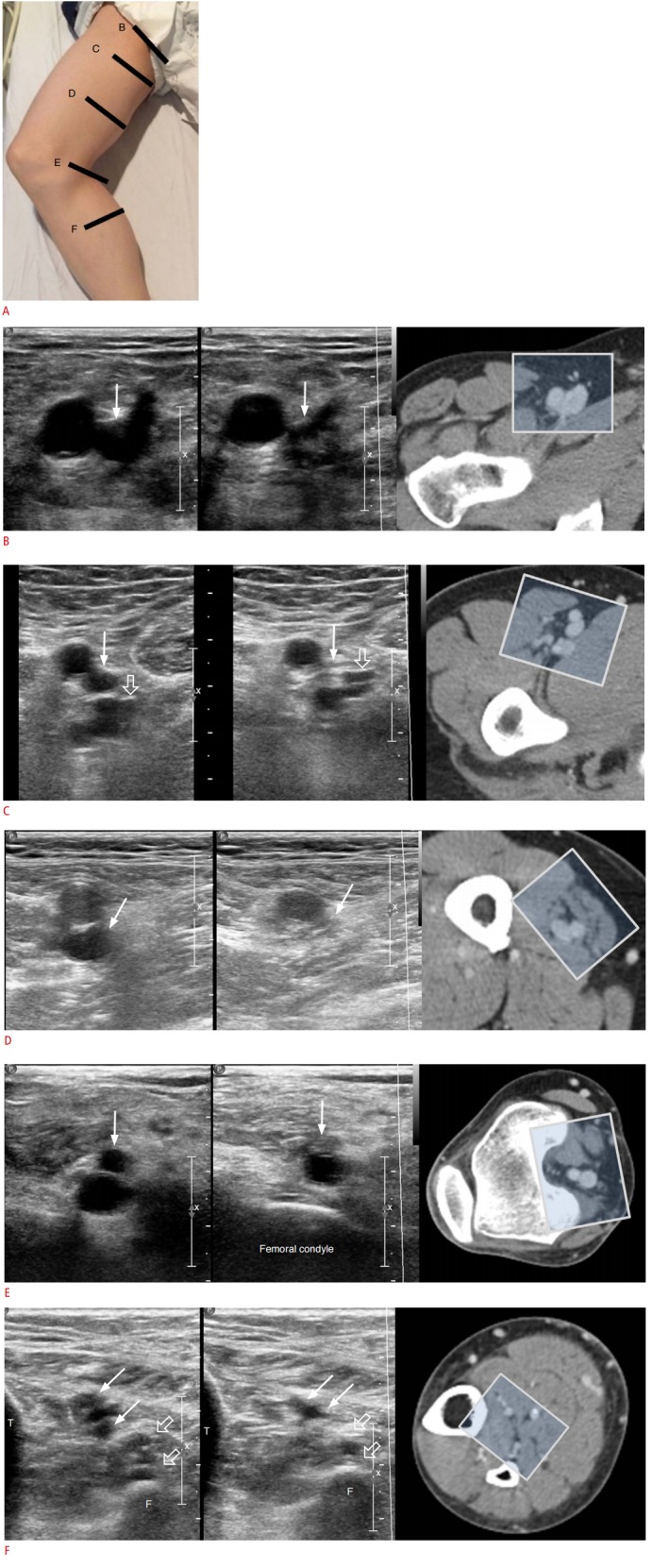
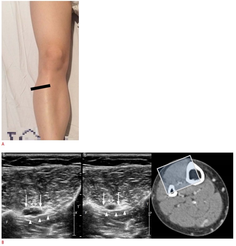
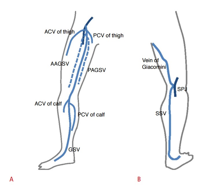
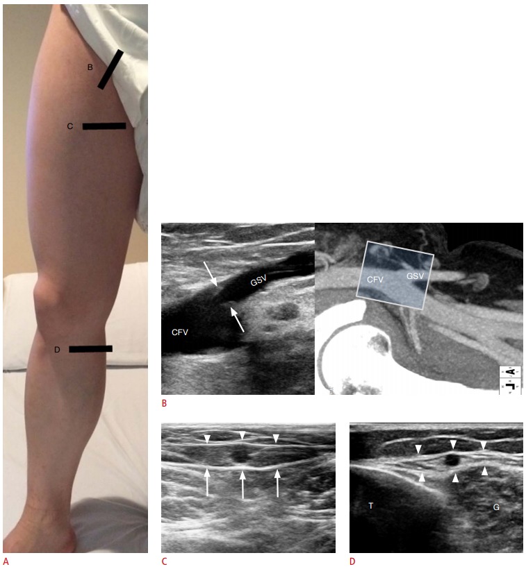
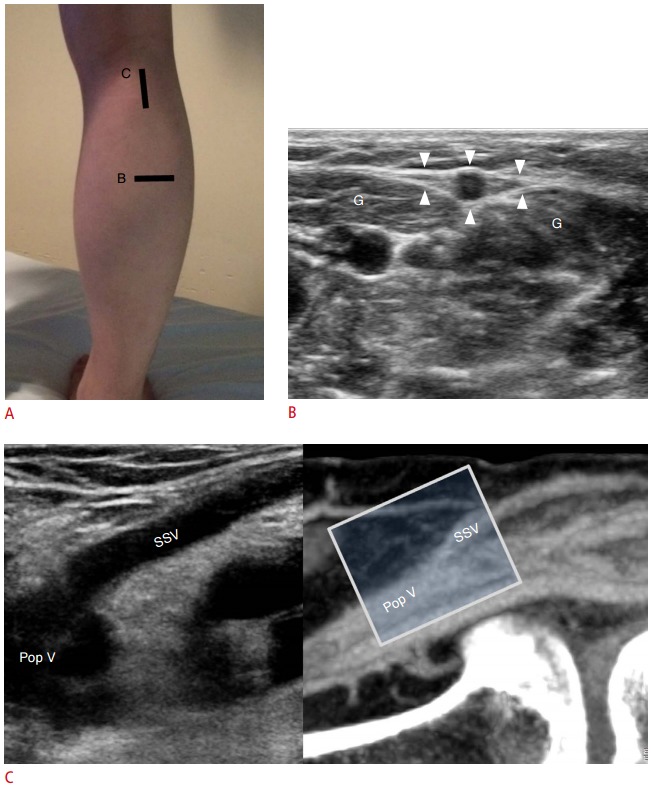
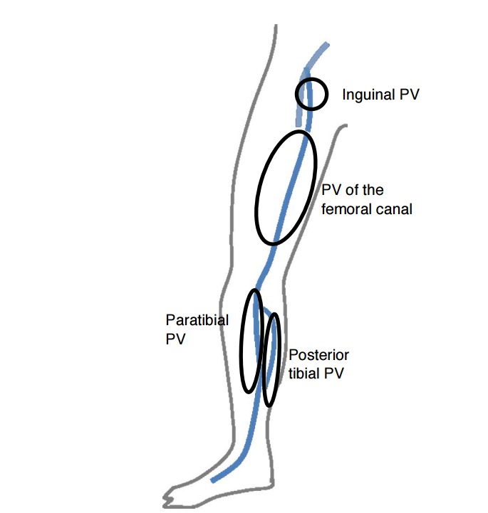
Ultrasonography is an imaging modality widely used to evaluate venous diseases of the lower extremities. It is important to understand the normal venous anatomy of the lower extremities, which has deep, superficial, and perforating venous components, in order to determine the pathophysiology of venous disease. This review provides a basic description of the anatomy of the lower extremity veins and useful techniques for approaching each vein via ultrasonography.
超声检查是一种广泛用于评估下肢静脉疾病的成像方式。了解下肢正常静脉解剖结构很重要,其具有深静脉、浅静脉和穿通静脉成分,以便确定静脉疾病的病理生理学。本综述提供了下肢静脉解剖结构的基本描述以及通过超声检查接近每条静脉的有用技术。