Hardbower D M, Coburn L A, Asim M, Singh K, Sierra J C, Barry D P, Gobert A P, Piazuelo M B, Washington M K, Wilson K T
Department of Pathology, Microbiology and Immunology, Vanderbilt University Medical Center, Nashville, TN, USA.
Division of Gastroenterology, Hepatology and Nutrition, Department of Medicine, Vanderbilt University Medical Center, Nashville, TN, USA.
Oncogene. 2017 Jul 6;36(27):3807-3819. doi: 10.1038/onc.2017.23. Epub 2017 Mar 6.
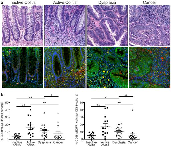
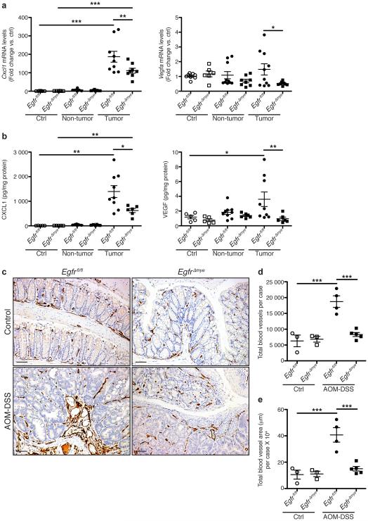
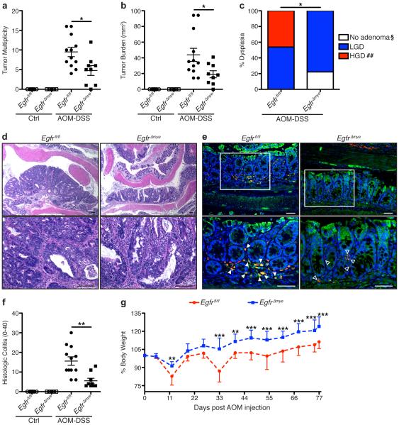
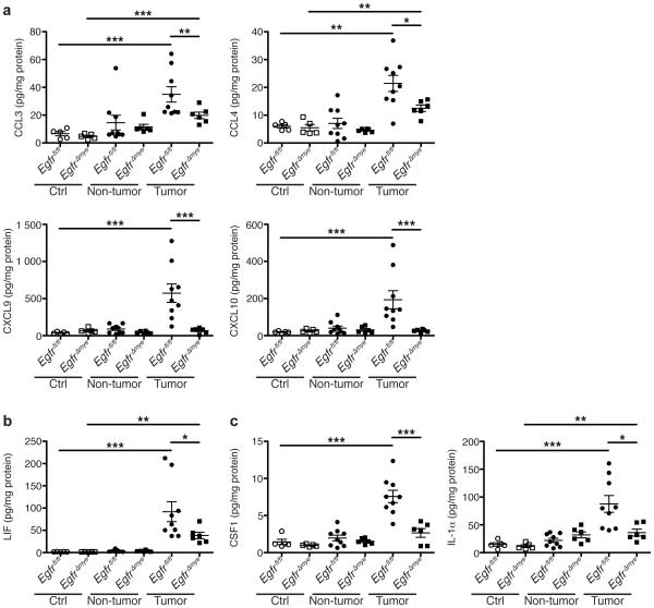
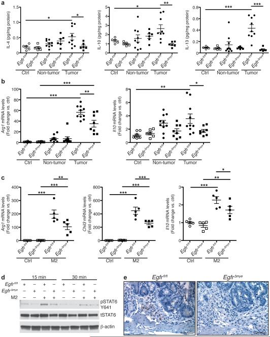
Epidermal growth factor receptor (EGFR) signaling is a known mediator of colorectal carcinogenesis. Studies have focused on the role of EGFR signaling in epithelial cells, although the exact nature of the role of EGFR in colorectal carcinogenesis remains a topic of debate. Here, we present evidence that EGFR signaling in myeloid cells, specifically macrophages, is critical for colon tumorigenesis in the azoxymethane-dextran sodium sulfate (AOM-DSS) model of colitis-associated carcinogenesis (CAC). In a human tissue microarray, colonic macrophages demonstrated robust EGFR activation in the pre-cancerous stages of colitis and dysplasia. Utilizing the AOM-DSS model, mice with a myeloid-specific deletion of Egfr had significantly decreased tumor multiplicity and burden, protection from high-grade dysplasia and significantly reduced colitis. Intriguingly, mice with gastrointestinal epithelial cell-specific Egfr deletion demonstrated no differences in tumorigenesis in the AOM-DSS model. The alterations in tumorigenesis in myeloid-specific Egfr knockout mice were accompanied by decreased macrophage, neutrophil and T-cell infiltration. Pro-tumorigenic M2 macrophage activation was diminished in myeloid-specific Egfr-deficient mice, as marked by decreased Arg1 and Il10 mRNA expression and decreased interleukin (IL)-4, IL10 and IL-13 protein levels. Surprisingly, diminished M1 macrophage activation was also detectable, as marked by significantly reduced Nos2 and Il1b mRNA levels and decreased interferon (IFN)-γ, tumor necrosis factor (TNF)-α and IL-1β protein levels. The alterations in M1 and M2 macrophage activation were confirmed in bone marrow-derived macrophages from mice with the myeloid-specific Egfr knockout. The combined effect of restrained M1 and M2 macrophage activation resulted in decreased production of pro-angiogenic factors, CXCL1 and vascular endothelial growth factor (VEGF), and reduced CD31 blood vessels, which likely contributed to protection from tumorigenesis. These data reveal that EGFR signaling in macrophages, but not in colonic epithelial cells, has a significant role in CAC. EGFR signaling in macrophages may prove to be an effective biomarker of CAC or target for chemoprevention in patients with inflammatory bowel disease.
表皮生长因子受体(EGFR)信号传导是已知的结直肠癌发生的介质。尽管EGFR在结直肠癌发生中的确切作用性质仍存在争议,但研究主要集中在EGFR信号传导在上皮细胞中的作用。在此,我们提供证据表明,在结肠炎相关癌(CAC)的氧化偶氮甲烷-葡聚糖硫酸钠(AOM-DSS)模型中,髓系细胞(特别是巨噬细胞)中的EGFR信号传导对结肠肿瘤发生至关重要。在人组织微阵列中,结肠巨噬细胞在结肠炎和发育异常的癌前阶段表现出强烈的EGFR激活。利用AOM-DSS模型,髓系特异性缺失Egfr的小鼠肿瘤数量和负担显著降低,对高级别发育异常具有保护作用,且结肠炎明显减轻。有趣的是,胃肠道上皮细胞特异性缺失Egfr的小鼠在AOM-DSS模型中的肿瘤发生没有差异。髓系特异性Egfr基因敲除小鼠肿瘤发生的改变伴随着巨噬细胞、中性粒细胞和T细胞浸润的减少。促肿瘤的M2巨噬细胞激活在髓系特异性Egfr缺陷小鼠中减弱,表现为Arg1和Il10 mRNA表达降低以及白细胞介素(IL)-4、IL10和IL-13蛋白水平降低。令人惊讶的是,也可检测到M1巨噬细胞激活减弱,表现为Nos2和Il1b mRNA水平显著降低以及干扰素(IFN)-γ、肿瘤坏死因子(TNF)-α和IL-1β蛋白水平降低。在髓系特异性Egfr基因敲除小鼠的骨髓来源巨噬细胞中证实了M1和M2巨噬细胞激活的改变。M1和M2巨噬细胞激活受到抑制的联合作用导致促血管生成因子CXCL1和血管内皮生长因子(VEGF)的产生减少以及CD31血管减少,这可能有助于预防肿瘤发生。这些数据表明,巨噬细胞而非结肠上皮细胞中的EGFR信号传导在CAC中具有重要作用。巨噬细胞中的EGFR信号传导可能被证明是CAC的有效生物标志物或炎症性肠病患者化学预防的靶点。