• 文献检索
  • 文档翻译
  • 深度研究
  • 学术资讯
  • Suppr Zotero 插件Zotero 插件
  • 邀请有礼
  • 套餐&价格
  • 历史记录
应用&插件
Suppr Zotero 插件Zotero 插件浏览器插件Mac 客户端Windows 客户端微信小程序
定价
高级版会员购买积分包购买API积分包
服务
文献检索文档翻译深度研究API 文档MCP 服务
关于我们
关于 Suppr公司介绍联系我们用户协议隐私条款
关注我们

Suppr 超能文献

核心技术专利:CN118964589B侵权必究
粤ICP备2023148730 号-1Suppr @ 2026

文献检索

告别复杂PubMed语法,用中文像聊天一样搜索,搜遍4000万医学文献。AI智能推荐,让科研检索更轻松。

立即免费搜索

文件翻译

保留排版,准确专业,支持PDF/Word/PPT等文件格式,支持 12+语言互译。

免费翻译文档

深度研究

AI帮你快速写综述,25分钟生成高质量综述,智能提取关键信息,辅助科研写作。

立即免费体验

利用结构张量同步辐射成像对心脏微观结构扩散张量磁共振成像测量进行验证。

Validation of diffusion tensor MRI measurements of cardiac microstructure with structure tensor synchrotron radiation imaging.

作者信息

Teh Irvin, McClymont Darryl, Zdora Marie-Christine, Whittington Hannah J, Davidoiu Valentina, Lee Jack, Lygate Craig A, Rau Christoph, Zanette Irene, Schneider Jürgen E

机构信息

Division of Cardiovascular Medicine, Radcliffe Department of Medicine, University of Oxford, Oxford, UK.

Leeds Institute of Cardiovascular & Metabolic Medicine, University of Leeds, Leeds, UK.

出版信息

J Cardiovasc Magn Reson. 2017 Mar 10;19(1):31. doi: 10.1186/s12968-017-0342-x.

DOI:10.1186/s12968-017-0342-x
PMID:28279178
原文链接:https://pmc.ncbi.nlm.nih.gov/articles/PMC5345150/
Abstract

BACKGROUND

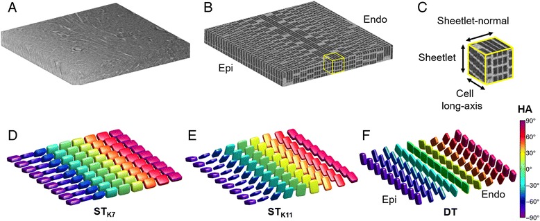
Diffusion tensor imaging (DTI) is widely used to assess tissue microstructure non-invasively. Cardiac DTI enables inference of cell and sheetlet orientations, which are altered under pathological conditions. However, DTI is affected by many factors, therefore robust validation is critical. Existing histological validation is intrinsically flawed, since it requires further tissue processing leading to sample distortion, is routinely limited in field-of-view and requires reconstruction of three-dimensional volumes from two-dimensional images. In contrast, synchrotron radiation imaging (SRI) data enables imaging of the heart in 3D without further preparation following DTI. The objective of the study was to validate DTI measurements based on structure tensor analysis of SRI data.

METHODS

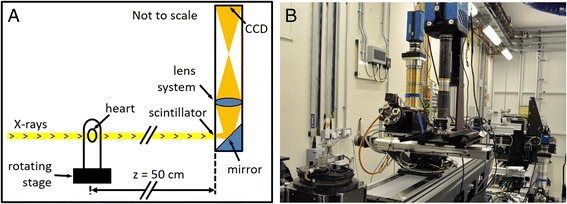
One isolated, fixed rat heart was imaged ex vivo with DTI and X-ray phase contrast SRI, and reconstructed at 100 μm and 3.6 μm isotropic resolution respectively. Structure tensors were determined from the SRI data and registered to the DTI data.

RESULTS

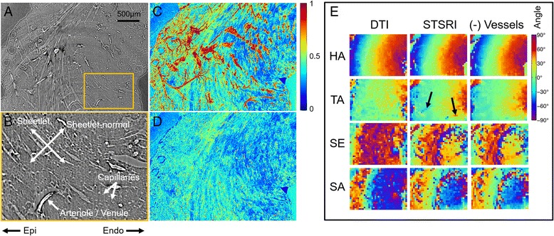
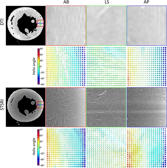
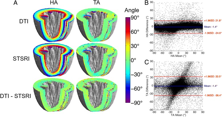
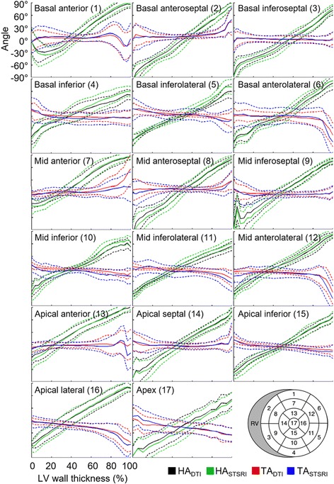
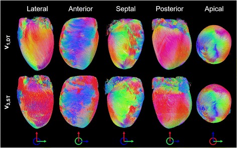
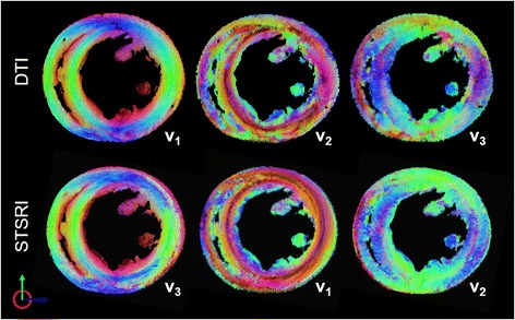
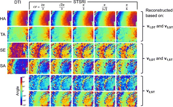
Excellent agreement in helix angles (HA) and transverse angles (TA) was observed between the DTI and structure tensor synchrotron radiation imaging (STSRI) data, where HA = -1.4° ± 23.2° and TA = -1.4° ± 35.0° (mean ± 1.96 standard deviation across all voxels in the left ventricle). STSRI confirmed that the primary eigenvector of the diffusion tensor corresponds with the cardiomyocyte long-axis across the whole myocardium.

CONCLUSIONS

We have used STSRI as a novel and high-resolution gold standard for the validation of DTI, allowing like-with-like comparison of three-dimensional tissue structures in the same intact heart free of distortion. This represents a critical step forward in independently verifying the structural basis and informing the interpretation of cardiac DTI data, thereby supporting the further development and adoption of DTI in structure-based electro-mechanical modelling and routine clinical applications.

摘要

背景

扩散张量成像(DTI)被广泛用于无创评估组织微观结构。心脏DTI能够推断细胞和心肌薄片的方向,这些方向在病理条件下会发生改变。然而,DTI受多种因素影响,因此可靠的验证至关重要。现有的组织学验证存在内在缺陷,因为它需要进一步的组织处理,这会导致样本变形,在视野上通常受到限制,并且需要从二维图像重建三维体积。相比之下,同步辐射成像(SRI)数据能够在DTI之后无需进一步准备即可对心脏进行三维成像。本研究的目的是基于SRI数据的结构张量分析来验证DTI测量结果。

方法

对一个离体固定的大鼠心脏进行体外DTI和X射线相衬SRI成像,并分别以100μm和3.6μm各向同性分辨率进行重建。从SRI数据中确定结构张量,并将其与DTI数据配准。

结果

在DTI和结构张量同步辐射成像(STSRI)数据之间观察到螺旋角(HA)和横向角(TA)具有极好的一致性,其中HA = -1.4°±23.2°,TA = -1.4°±35.0°(左心室所有体素的平均值±1.96标准差)。STSRI证实,扩散张量的主特征向量与整个心肌中的心肌细胞长轴相对应。

结论

我们已将STSRI用作验证DTI的一种新颖且高分辨率的金标准,能够在同一完整且无变形的心脏中对三维组织结构进行同类比较。这代表了在独立验证结构基础并为心脏DTI数据的解释提供依据方面向前迈出的关键一步,从而支持DTI在基于结构的机电建模和常规临床应用中的进一步发展和应用。

https://cdn.ncbi.nlm.nih.gov/pmc/blobs/e6be/5345150/615c92b9d533/12968_2017_342_Fig9_HTML.jpg
https://cdn.ncbi.nlm.nih.gov/pmc/blobs/e6be/5345150/396c96b3f6b8/12968_2017_342_Fig1_HTML.jpg
https://cdn.ncbi.nlm.nih.gov/pmc/blobs/e6be/5345150/44abcd0d8bdd/12968_2017_342_Fig2_HTML.jpg
https://cdn.ncbi.nlm.nih.gov/pmc/blobs/e6be/5345150/977060a4d536/12968_2017_342_Fig3_HTML.jpg
https://cdn.ncbi.nlm.nih.gov/pmc/blobs/e6be/5345150/5bb37fe47c36/12968_2017_342_Fig4_HTML.jpg
https://cdn.ncbi.nlm.nih.gov/pmc/blobs/e6be/5345150/c45464a1eb88/12968_2017_342_Fig5_HTML.jpg
https://cdn.ncbi.nlm.nih.gov/pmc/blobs/e6be/5345150/1e9169108fed/12968_2017_342_Fig6_HTML.jpg
https://cdn.ncbi.nlm.nih.gov/pmc/blobs/e6be/5345150/00323809d0fc/12968_2017_342_Fig7_HTML.jpg
https://cdn.ncbi.nlm.nih.gov/pmc/blobs/e6be/5345150/604823a6c71b/12968_2017_342_Fig8_HTML.jpg
https://cdn.ncbi.nlm.nih.gov/pmc/blobs/e6be/5345150/615c92b9d533/12968_2017_342_Fig9_HTML.jpg

相似文献

1
Validation of diffusion tensor MRI measurements of cardiac microstructure with structure tensor synchrotron radiation imaging.利用结构张量同步辐射成像对心脏微观结构扩散张量磁共振成像测量进行验证。
J Cardiovasc Magn Reson. 2017 Mar 10;19(1):31. doi: 10.1186/s12968-017-0342-x.
2
Comparison of diffusion tensor imaging by cardiovascular magnetic resonance and gadolinium enhanced 3D image intensity approaches to investigation of structural anisotropy in explanted rat hearts.心血管磁共振扩散张量成像与钆增强三维图像强度法在离体大鼠心脏结构各向异性研究中的比较
J Cardiovasc Magn Reson. 2015 Apr 29;17(1):31. doi: 10.1186/s12968-015-0129-x.
3
The impact of signal-to-noise ratio, diffusion-weighted directions and image resolution in cardiac diffusion tensor imaging - insights from the ex-vivo rat heart.信噪比、扩散加权方向和图像分辨率对心脏扩散张量成像的影响——来自离体大鼠心脏的观察。
J Cardiovasc Magn Reson. 2017 Nov 20;19(1):90. doi: 10.1186/s12968-017-0395-x.
4
Whole heart detailed and quantitative anatomy, myofibre structure and vasculature from X-ray phase-contrast synchrotron radiation-based micro computed tomography.基于同步辐射 X 射线相衬的微计算机断层扫描的全心详细定量解剖、肌纤维结构和脉管系统。
Eur Heart J Cardiovasc Imaging. 2017 Jul 1;18(7):732-741. doi: 10.1093/ehjci/jew314.
5
Accelerating cardiac diffusion tensor imaging combining local low-rank and 3D TV constraint.加速心脏扩散张量成像的局部低秩和 3D TV 约束相结合。
MAGMA. 2019 Aug;32(4):407-422. doi: 10.1007/s10334-019-00747-1. Epub 2019 Mar 22.
6
A high-resolution cardiovascular magnetic resonance diffusion tensor map from ex-vivo C57BL/6 murine hearts.来自离体C57BL/6小鼠心脏的高分辨率心血管磁共振扩散张量图。
J Cardiovasc Magn Reson. 2014 Oct 16;16(1):77. doi: 10.1186/s12968-014-0077-x.
7
Microarchitecture of the hearts in term and former-preterm lambs using diffusion tensor imaging.使用扩散张量成像技术研究足月和早产羔羊心脏的微观结构。
Anat Rec (Hoboken). 2021 Apr;304(4):803-817. doi: 10.1002/ar.24516. Epub 2020 Oct 20.
8
Adaptive anisotropic gaussian filtering to reduce acquisition time in cardiac diffusion tensor imaging.自适应各向异性高斯滤波以减少心脏扩散张量成像中的采集时间。
Int J Cardiovasc Imaging. 2016 Jun;32(6):921-34. doi: 10.1007/s10554-016-0848-6. Epub 2016 Feb 2.
9
Three-dimensional Cardiomyocytes Structure Revealed By Diffusion Tensor Imaging and Its Validation Using a Tissue-Clearing Technique.三维心肌细胞结构的扩散张量成像揭示及其使用组织透明化技术的验证。
Sci Rep. 2018 Apr 27;8(1):6640. doi: 10.1038/s41598-018-24622-6.
10
Direct comparison of in vivo versus postmortem second-order motion-compensated cardiac diffusion tensor imaging.体内与死后的二阶运动补偿心脏扩散张量成像的直接比较。
Magn Reson Med. 2018 Apr;79(4):2265-2276. doi: 10.1002/mrm.26871. Epub 2017 Aug 22.

引用本文的文献

1
Cardiac diffusion-weighted and tensor imaging: A consensus statement from the special interest group of the Society for Cardiovascular Magnetic Resonance.心脏扩散加权成像和张量成像:心血管磁共振学会特别兴趣小组的共识声明
J Cardiovasc Magn Reson. 2024 Oct 22;27(1):101109. doi: 10.1016/j.jocmr.2024.101109.
2
The effects of field strength on stimulated echo and motion-compensated spin-echo diffusion tensor cardiovascular magnetic resonance sequences.场强对受激回波和运动补偿自旋回波扩散张量心血管磁共振序列的影响。
J Cardiovasc Magn Reson. 2024;26(2):101052. doi: 10.1016/j.jocmr.2024.101052. Epub 2024 Jun 25.
3

本文引用的文献

1
Resolving Fine Cardiac Structures in Rats with High-Resolution Diffusion Tensor Imaging.利用高分辨率扩散张量成像解析大鼠精细心脏结构
Sci Rep. 2016 Jul 28;6:30573. doi: 10.1038/srep30573.
2
Mapping cardiac microstructure of rabbit heart in different mechanical states by high resolution diffusion tensor imaging: A proof-of-principle study.通过高分辨率扩散张量成像绘制不同力学状态下兔心脏的心脏微观结构:一项原理验证研究。
Prog Biophys Mol Biol. 2016 Jul;121(2):85-96. doi: 10.1016/j.pbiomolbio.2016.06.001. Epub 2016 Jun 16.
3
Cardiac Light-Sheet Fluorescent Microscopy for Multi-Scale and Rapid Imaging of Architecture and Function.
Cardiomyocyte orientation recovery at micrometer scale reveals long-axis fiber continuum in heart walls.
心肌细胞在微米尺度上的取向恢复揭示了心壁中长轴纤维的连续性。
EMBO J. 2023 Oct 4;42(19):e113288. doi: 10.15252/embj.2022113288. Epub 2023 Sep 6.
4
Measuring cardiomyocyte cellular characteristics in cardiac hypertrophy using diffusion-weighted MRI.应用扩散加权 MRI 测量心肌肥厚中的心肌细胞特征。
Magn Reson Med. 2023 Nov;90(5):2144-2157. doi: 10.1002/mrm.29775. Epub 2023 Jun 22.
5
Effects of myocardial sheetlet sliding on left ventricular function.心肌片滑动对左心室功能的影响。
Biomech Model Mechanobiol. 2023 Aug;22(4):1313-1332. doi: 10.1007/s10237-023-01721-6. Epub 2023 May 6.
6
The relationship between myocardial microstructure and strain in chronic infarction using cardiovascular magnetic resonance diffusion tensor imaging and feature tracking.应用心血管磁共振弥散张量成像和特征追踪技术研究慢性梗死心肌微结构与应变的关系。
J Cardiovasc Magn Reson. 2022 Nov 24;24(1):66. doi: 10.1186/s12968-022-00892-y.
7
A groupwise registration and tractography framework for cardiac myofiber architecture description by diffusion MRI: An application to the ventricular junctions.基于扩散 MRI 的心肌纤维结构描述的分组配准和束追踪框架:在心室交界处的应用。
PLoS One. 2022 Jul 18;17(7):e0271279. doi: 10.1371/journal.pone.0271279. eCollection 2022.
8
Myocardial mesostructure and mesofunction.心肌中结构和中功能。
Am J Physiol Heart Circ Physiol. 2022 Aug 1;323(2):H257-H275. doi: 10.1152/ajpheart.00059.2022. Epub 2022 Jun 3.
9
In Vivo Super-Resolution Cardiac Diffusion Tensor MRI: A Feasibility Study.体内超分辨率心脏扩散张量磁共振成像:一项可行性研究。
Diagnostics (Basel). 2022 Mar 31;12(4):877. doi: 10.3390/diagnostics12040877.
10
Impact of intraventricular septal fiber orientation on cardiac electromechanical function.室间隔纤维方向对心机电功能的影响。
Am J Physiol Heart Circ Physiol. 2022 Jun 1;322(6):H936-H952. doi: 10.1152/ajpheart.00050.2022. Epub 2022 Mar 18.
用于心脏结构与功能多尺度快速成像的心脏光片荧光显微镜技术
Sci Rep. 2016 Mar 3;6:22489. doi: 10.1038/srep22489.
4
Contrast-free detection of myocardial fibrosis in hypertrophic cardiomyopathy patients with diffusion-weighted cardiovascular magnetic resonance.利用扩散加权心血管磁共振对肥厚型心肌病患者进行无对比剂心肌纤维化检测。
J Cardiovasc Magn Reson. 2015 Dec 2;17:107. doi: 10.1186/s12968-015-0214-1.
5
Ultra-high-resolution 3D imaging of atherosclerosis in mice with synchrotron differential phase contrast: a proof of concept study.同步辐射微分相衬成像对小鼠动脉粥样硬化的超高分辨率三维成像:一项概念验证研究。
Sci Rep. 2015 Jul 13;5:11980. doi: 10.1038/srep11980.
6
Comparison of diffusion tensor imaging by cardiovascular magnetic resonance and gadolinium enhanced 3D image intensity approaches to investigation of structural anisotropy in explanted rat hearts.心血管磁共振扩散张量成像与钆增强三维图像强度法在离体大鼠心脏结构各向异性研究中的比较
J Cardiovasc Magn Reson. 2015 Apr 29;17(1):31. doi: 10.1186/s12968-015-0129-x.
7
Higher-Order Motion-Compensation for In Vivo Cardiac Diffusion Tensor Imaging in Rats.大鼠体内心脏扩散张量成像的高阶运动补偿
IEEE Trans Med Imaging. 2015 Sep;34(9):1843-53. doi: 10.1109/TMI.2015.2411571. Epub 2015 Mar 9.
8
In vivo cardiovascular magnetic resonance diffusion tensor imaging shows evidence of abnormal myocardial laminar orientations and mobility in hypertrophic cardiomyopathy.体内心血管磁共振扩散张量成像显示肥厚型心肌病中心肌层状取向和运动异常的证据。
J Cardiovasc Magn Reson. 2014 Nov 12;16(1):87. doi: 10.1186/s12968-014-0087-8.
9
In vivo contrast free chronic myocardial infarction characterization using diffusion-weighted cardiovascular magnetic resonance.使用扩散加权心血管磁共振对体内无对比剂的慢性心肌梗死进行特征描述。
J Cardiovasc Magn Reson. 2014 Sep 17;16(1):68. doi: 10.1186/s12968-014-0068-y.
10
Interrogation of living myocardium in multiple static deformation states with diffusion tensor and diffusion spectrum imaging.利用扩散张量成像和扩散谱成像对处于多种静态变形状态的活体心肌进行检测。
Prog Biophys Mol Biol. 2014 Aug;115(2-3):213-25. doi: 10.1016/j.pbiomolbio.2014.08.002. Epub 2014 Aug 10.