Moulisová Vladimíra, Gonzalez-García Cristina, Cantini Marco, Rodrigo-Navarro Aleixandre, Weaver Jessica, Costell Mercedes, Sabater I Serra Roser, Dalby Matthew J, García Andrés J, Salmerón-Sánchez Manuel
Division of Biomedical Engineering, School of Engineering, University of Glasgow, Glasgow G12 8LT, United Kingdom.
Woodruff School of Mechanical Engineering, Petit Institute for Bioengineering and Bioscience, Georgia Institute of Technology, Atlanta, GA 30332, USA.
Biomaterials. 2017 May;126:61-74. doi: 10.1016/j.biomaterials.2017.02.024. Epub 2017 Feb 21.
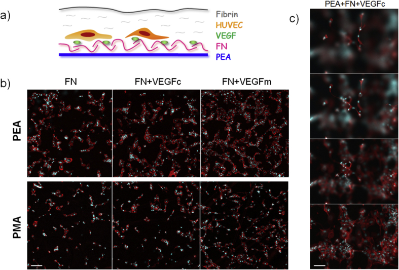
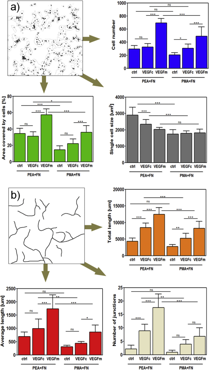
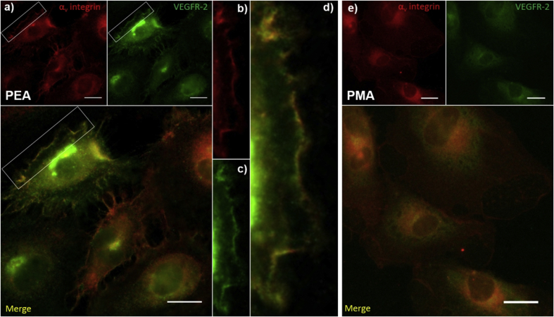
We have engineered polymer-based microenvironments that promote vasculogenesis both in vitro and in vivo through synergistic integrin-growth factor receptor signalling. Poly(ethyl acrylate) (PEA) triggers spontaneous organization of fibronectin (FN) into nanonetworks which provide availability of critical binding domains. Importantly, the growth factor binding (FNIII) and integrin binding (FNIII) regions are simultaneously available on FN fibrils assembled on PEA. This material platform promotes synergistic integrin/VEGF signalling which is highly effective for vascularization events in vitro with low concentrations of VEGF. VEGF specifically binds to FN fibrils on PEA compared to control polymers (poly(methyl acrylate), PMA) where FN remains in a globular conformation and integrin/GF binding domains are not simultaneously available. The vasculogenic response of human endothelial cells seeded on these synergistic interfaces (VEGF bound to FN assembled on PEA) was significantly improved compared to soluble administration of VEGF at higher doses. Early onset of VEGF signalling (PLCγ1 phosphorylation) and both integrin and VEGF signalling (ERK1/2 phosphorylation) were increased only when VEGF was bound to FN nanonetworks on PEA, while soluble VEGF did not influence early signalling. Experiments with mutant FN molecules with impaired integrin binding site (FN-RGE) confirmed the role of the integrin binding site of FN on the vasculogenic response via combined integrin/VEGF signalling. In vivo experiments using 3D scaffolds coated with FN and VEGF implanted in the murine fat pad demonstrated pro-vascularization signalling by enhanced formation of new tissue inside scaffold pores. PEA-driven organization of FN promotes efficient presentation of VEGF to promote vascularization in regenerative medicine applications.
我们构建了基于聚合物的微环境,通过整合素 - 生长因子受体信号协同作用,在体外和体内促进血管生成。聚(丙烯酸乙酯)(PEA)触发纤连蛋白(FN)自发组织形成纳米网络,提供关键结合域的可用性。重要的是,在PEA上组装的FN原纤维上,生长因子结合(FNIII)和整合素结合(FNIII)区域同时可用。这个材料平台促进整合素/VEGF信号协同作用,对于低浓度VEGF的体外血管化事件非常有效。与对照聚合物(聚(甲基丙烯酸甲酯),PMA)相比,VEGF特异性结合PEA上的FN原纤维,在对照聚合物中FN保持球状构象,整合素/生长因子结合域不同时可用。与高剂量可溶性VEGF给药相比,接种在这些协同界面(VEGF结合到PEA上组装的FN)上的人内皮细胞的血管生成反应显著改善。仅当VEGF结合到PEA上的FN纳米网络时,VEGF信号的早期启动(PLCγ1磷酸化)以及整合素和VEGF信号(ERK1/2磷酸化)才会增加,而可溶性VEGF不影响早期信号。使用整合素结合位点受损的突变FN分子(FN-RGE)进行的实验证实了FN的整合素结合位点通过整合素/VEGF信号组合对血管生成反应的作用。使用涂有FN和VEGF的3D支架植入小鼠脂肪垫的体内实验表明,通过支架孔内新组织的增强形成促进血管化信号。PEA驱动的FN组织促进VEGF的有效呈递,以促进再生医学应用中的血管化。