Llopis-Hernández Virginia, Rico Patricia, Moratal David, Altankov George, Salmerón-Sánchez Manuel
Center for Biomaterials and Tissue Engineering, Universitat Politècnica de València , Valencia, Spain .
Biores Open Access. 2013 Oct;2(5):364-73. doi: 10.1089/biores.2013.0017.
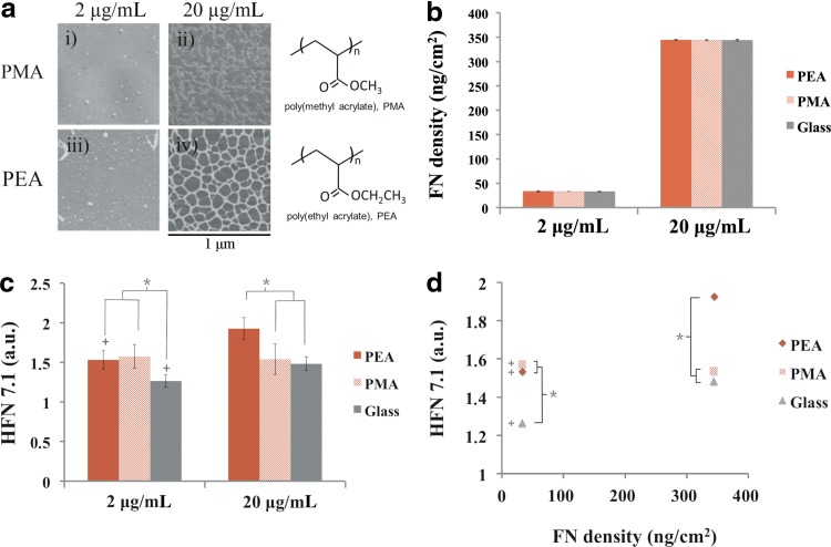
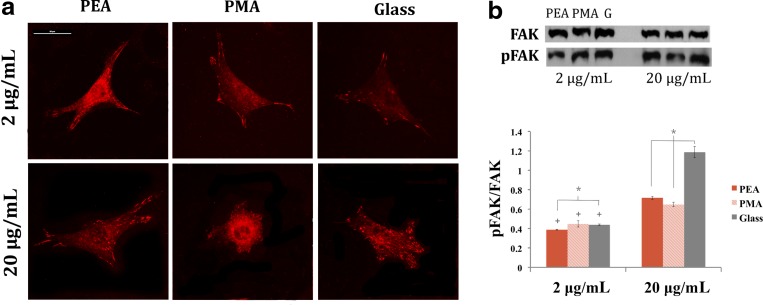
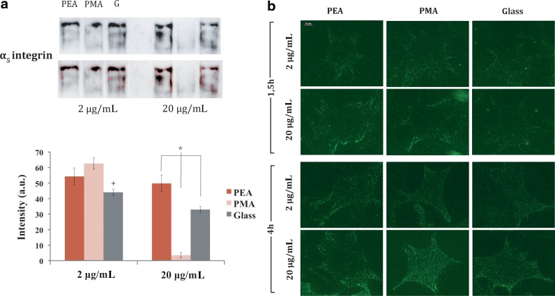
Protein remodeling at the cell-material interface is an important phenomenon that should be incorporated into the design of advanced biomaterials for tissue engineering. In this work, we address the relationship between fibronectin (FN) activity at the material interface and remodeling, including proteolytic cascades. To do so, we studied FN adsorption on two chemically similar substrates, poly(ethyl acrylate) (PEA) and poly(methyl acrylate) (PMA), which resulted in different distribution and conformation of the protein at the material interface: FN organized spontaneously upon adsorption on PEA into physiological-like fibrils, through a process called material-driven FN fibrillogenesis. The amount of adsorbed FN and its conformation were investigated in two different coating concentrations (2 and 20 μg/mL). Since FN activity at the material interface determines the initial cellular response, we followed the formation of focal adhesions (vinculin) and subsequent cell signaling by focal adhesion kinase (FAK) expression and its phosphorylation (pFAK). More detailed studies were performed to get further insights into integrin binding by crosslinking and extraction followed by immunofluorescence, as well as protein and gene expression for α5 and αv. To correlate cell adhesion with matrix degradation, gene expression and activity (zymography) of matrix metalloproteinases (MMPs) were measured. Overall, we demonstrated that the material-driven FN fibrillogenesis triggers proteolytic activity: MMP activity was higher on the material-driven FN fibrils, as a compensatory mechanism to the inability of cells to reorganize this FN network.
细胞与材料界面处的蛋白质重塑是一种重要现象,应纳入用于组织工程的先进生物材料设计中。在这项工作中,我们研究了材料界面处纤连蛋白(FN)活性与重塑之间的关系,包括蛋白水解级联反应。为此,我们研究了FN在两种化学性质相似的底物聚(丙烯酸乙酯)(PEA)和聚(丙烯酸甲酯)(PMA)上的吸附情况,这导致蛋白质在材料界面处具有不同的分布和构象:FN在吸附到PEA上时通过一种称为材料驱动的FN原纤维形成过程自发组织成类似生理状态的原纤维。在两种不同的包被浓度(2和20μg/mL)下研究了吸附的FN量及其构象。由于材料界面处的FN活性决定了最初的细胞反应,我们通过粘着斑(纽蛋白)的形成以及粘着斑激酶(FAK)表达及其磷酸化(pFAK)来追踪后续的细胞信号传导。通过交联和提取后进行免疫荧光以及α5和αv的蛋白质和基因表达,进行了更详细的研究以进一步了解整合素结合情况。为了将细胞粘附与基质降解相关联,测量了基质金属蛋白酶(MMPs)的基因表达和活性(酶谱分析)。总体而言,我们证明了材料驱动的FN原纤维形成触发了蛋白水解活性:MMP活性在材料驱动的FN原纤维上更高,作为对细胞无法重组该FN网络的一种补偿机制。