Papma Janne M, Smits Marion, de Groot Marius, Mattace Raso Francesco U, van der Lugt Aad, Vrooman Henri A, Niessen Wiro J, Koudstaal Peter J, van Swieten John C, van der Veen Frederik M, Prins Niels D
Department of Neurology, Erasmus MC - University Medical Center Rotterdam, 's-Gravendijkwal 230, 3015 CE, Rotterdam, The Netherlands.
Department of Radiology, Erasmus MC - University Medical Center Rotterdam, Rotterdam, The Netherlands.
Eur Radiol. 2017 Sep;27(9):3716-3724. doi: 10.1007/s00330-017-4768-1. Epub 2017 Mar 13.
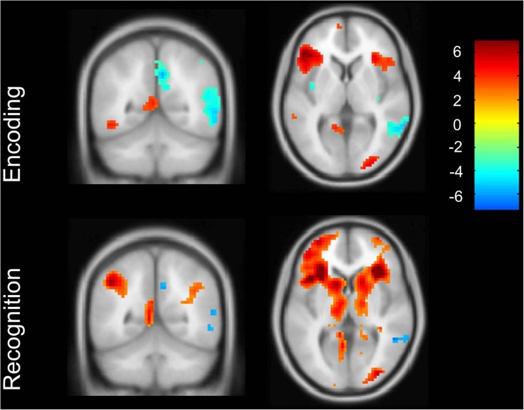
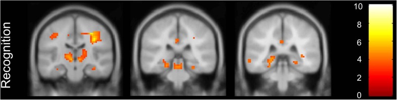
Diminished function of the posterior cingulate cortex (PCC) is a typical finding in early Alzheimer's disease (AD). It is hypothesized that in early stage AD, PCC functioning relates to or reflects hippocampal dysfunction or atrophy. The aim of this study was to examine the relationship between hippocampus function, volume and structural connectivity, and PCC activation during an episodic memory task-related fMRI study in mild cognitive impairment (MCI).
MCI patients (n = 27) underwent episodic memory task-related fMRI, 3D-T1w MRI, 2D T2-FLAIR MRI and diffusion tensor imaging. Stepwise linear regression analysis was performed to examine the relationship between PCC activation and hippocampal activation, hippocampal volume and diffusion measures within the cingulum along the hippocampus.
We found a significant relationship between PCC and hippocampus activation during successful episodic memory encoding and correct recognition in MCI patients. We found no relationship between the PCC and structural hippocampal predictors.
Our results indicate a relationship between PCC and hippocampus activation during episodic memory engagement in MCI. This may suggest that during episodic memory, functional network deterioration is the most important predictor of PCC functioning in MCI.
• PCC functioning during episodic memory relates to hippocampal functioning in MCI. • PCC functioning during episodic memory does not relate to hippocampal structure in MCI. • Functional network changes are an important predictor of PCC functioning in MCI.
后扣带回皮质(PCC)功能减退是早期阿尔茨海默病(AD)的典型表现。据推测,在AD早期,PCC功能与海马功能障碍或萎缩有关或反映了海马功能障碍或萎缩。本研究的目的是在轻度认知障碍(MCI)患者的情景记忆任务相关功能磁共振成像(fMRI)研究中,检验海马功能、体积和结构连接性与PCC激活之间的关系。
MCI患者(n = 27)接受了情景记忆任务相关fMRI、三维T1加权磁共振成像(3D-T1w MRI)、二维液体衰减反转恢复序列磁共振成像(2D T2-FLAIR MRI)和扩散张量成像。进行逐步线性回归分析,以检验PCC激活与海马激活、海马体积以及沿海马的扣带束扩散测量值之间的关系。
我们发现,在MCI患者成功进行情景记忆编码和正确识别期间,PCC与海马激活之间存在显著关系。我们发现PCC与海马结构预测指标之间没有关系。
我们的结果表明,在MCI患者进行情景记忆时,PCC与海马激活之间存在关系。这可能表明,在情景记忆过程中,功能网络退化是MCI中PCC功能的最重要预测指标。
• MCI患者情景记忆期间的PCC功能与海马功能有关。• MCI患者情景记忆期间的PCC功能与海马结构无关。• 功能网络变化是MCI中PCC功能的重要预测指标。