Pennanen Paula, Alanne Maria Helena, Fazeli Elnaz, Deguchi Takahiro, Näreoja Tuomas, Peltonen Sirkku, Peltonen Juha
Department of Cell Biology and Anatomy, Institute of Biomedicine, University of Turku, Kiinamyllynkatu 10, 20520, Turku, Finland.
Laboratory of Biophysics, Department of Cell Biology and Anatomy and Medicity Research Laboratories, University of Turku, P.O. Box 123, 20521, Turku, Finland.
Mol Cell Biochem. 2017 Aug;432(1-2):131-139. doi: 10.1007/s11010-017-3004-2. Epub 2017 Mar 14.
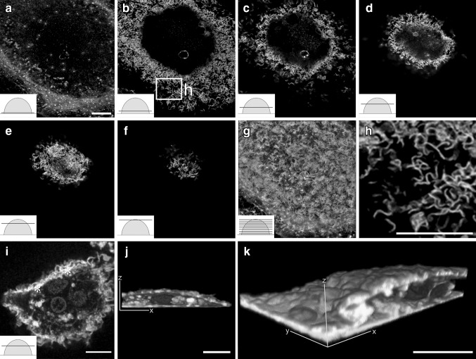
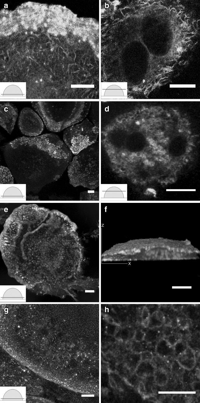
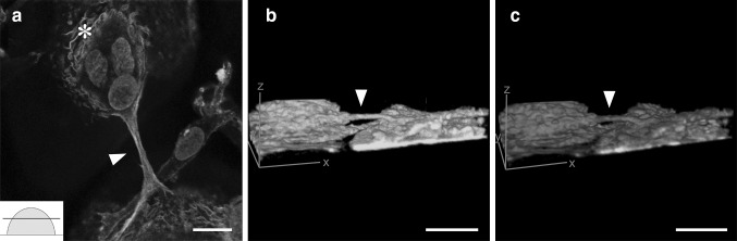
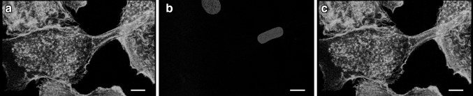
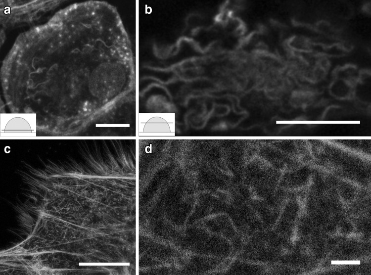
Osteoclasts are multinucleated bone-resorbing cells with a dynamic actin cytoskeleton. Osteoclasts are derived from circulating mononuclear precursors. Confocal and stimulated emission depletion (STED) super-resolution microscopy was used to investigate peripheral blood-derived human osteoclasts cultured on glass surfaces. STED and confocal microscopy demonstrated that the actin was curved and branched, for instance, in the vicinity of membrane ruffles. The overall architecture of the curved actin network extended from the podosomes to the top of the cell. The other novel finding was that a micrometer-level tube containing actin bridged the osteoclasts well above the level of the culture glass. The actin filaments of the tubes originated from the network of curved actin often surrounding a group of nuclei. Furthermore, nuclei were occasionally located inside the tubes. Our findings demonstrated the accumulation of c-Src, cortactin, cofilin, and actin around nuclei suggesting their role in nuclear processes such as the locomotion of nuclei. ARP2/3 labeling was abundant at the substratum level of osteoclasts and in the branched actin network, where it localized to the branching points. We speculate that the actin-containing tubes of osteoclasts may provide a means of transportation of nuclei, e.g., during the fusion of osteoclasts. These novel findings can pave the way for future studies aiming at the elucidation of the differentiation of multinucleated osteoclasts.
破骨细胞是具有动态肌动蛋白细胞骨架的多核骨吸收细胞。破骨细胞来源于循环单核前体细胞。利用共聚焦和受激发射损耗(STED)超分辨率显微镜研究在玻璃表面培养的外周血来源的人破骨细胞。STED和共聚焦显微镜显示,例如在膜皱褶附近,肌动蛋白呈弯曲和分支状。弯曲的肌动蛋白网络的整体结构从足体延伸到细胞顶部。另一个新发现是,一个含有肌动蛋白的微米级管道在培养玻璃平面上方连接着破骨细胞。管道的肌动蛋白丝起源于通常围绕一组细胞核的弯曲肌动蛋白网络。此外,细胞核偶尔位于管道内部。我们的研究结果表明,c-Src、皮层肌动蛋白、丝切蛋白和肌动蛋白在细胞核周围积累,表明它们在细胞核运动等核过程中发挥作用。ARP2/3标记在破骨细胞的基质水平和分支的肌动蛋白网络中丰富,其定位于分支点。我们推测,破骨细胞含肌动蛋白的管道可能提供一种细胞核运输方式,例如在破骨细胞融合过程中。这些新发现可为今后旨在阐明多核破骨细胞分化的研究铺平道路。