Ma Liang, Li Ming-Wei, Bai Yu, Guo Hui-Hui, Wang Sheng-Chao, Yu Qing
State Key Laboratory of Military Stomatology & National Clinical Research Center for Oral Diseases & Shaanxi Key Laboratory of Oral Diseases, Department of Operative Dentistry and Endodontics, The Fourth Military Medical University, Xi'an, China; Department of Stomatology, No. 44 Hospital of Chinese PLA, Guiyang, Guizhou, China.
State Key Laboratory of Military Stomatology & National Clinical Research Center for Oral Diseases & Shaanxi Key Laboratory of Oral Diseases, Department of Operative Dentistry and Endodontics, The Fourth Military Medical University, Xi'an, China.
Stem Cells Int. 2017;2017:4837503. doi: 10.1155/2017/4837503. Epub 2017 Feb 16.
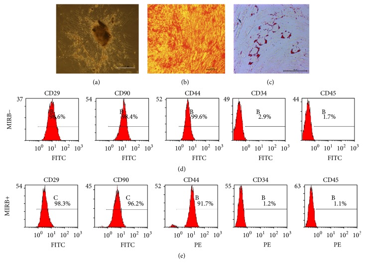
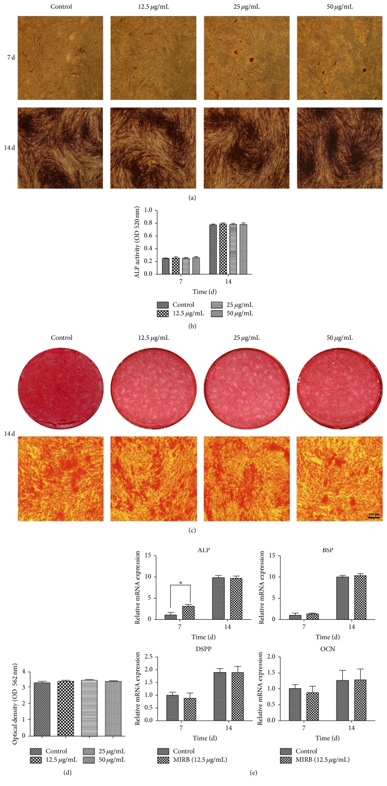
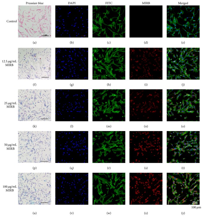
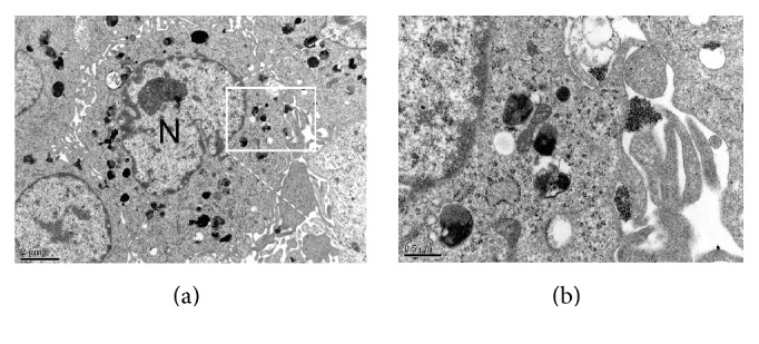
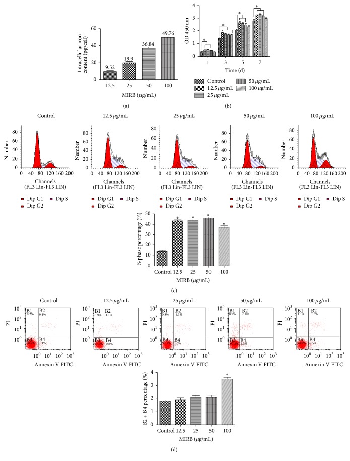
Tracking transplanted stem cells is necessary to clarify cellular properties and improve transplantation success. In this study, we investigate the effects of fluorescent superparamagnetic iron oxide particles (SPIO) (Molday ION Rhodamine-B™, MIRB) on biological properties of human dental pulp stem cells (hDPSCs) and monitor hDPSCs in vitro and in vivo using magnetic resonance imaging (MRI). Morphological analysis showed that intracellular MIRB particles were distributed in the cytoplasm surrounding the nuclei of hDPSCs. 12.5-100 g/mL MIRB all resulted in 100% labeling efficiency. MTT showed that 12.5-50 g/mL MIRB could promote cell proliferation and MIRB over 100 g/mL exhibited toxic effect on hDPSCs. In vitro MRI showed that 1 × 10 cells labeled with various concentrations of MIRB (12.5-100 g/mL) could be visualized. In vivo MRI showed that transplanted cells could be clearly visualized up to 60 days after transplantation. These results suggest that 12.5-50 g/mL MIRB is a safe range for labeling hDPSCs. MIRB labeled hDPSCs cell can be visualized by MRI in vitro and in vivo. These data demonstrate that MIRB is a promising candidate for hDPSCs tracking in hDPSCs based dental pulp regeneration therapy.
追踪移植的干细胞对于阐明细胞特性和提高移植成功率至关重要。在本研究中,我们研究了荧光超顺磁性氧化铁颗粒(SPIO)(莫尔代离子罗丹明 - B™,MIRB)对人牙髓干细胞(hDPSCs)生物学特性的影响,并使用磁共振成像(MRI)在体外和体内监测hDPSCs。形态学分析表明,细胞内的MIRB颗粒分布在hDPSCs细胞核周围的细胞质中。12.5 - 100μg/mL的MIRB均导致100%的标记效率。MTT法显示,12.5 - 50μg/mL的MIRB可促进细胞增殖,而超过100μg/mL的MIRB对hDPSCs表现出毒性作用。体外MRI显示,用不同浓度(12.5 - 100μg/mL)的MIRB标记的1×10⁶个细胞可以被可视化。体内MRI显示,移植后长达60天均可清晰地观察到移植的细胞。这些结果表明,12.5 - 50μg/mL的MIRB是标记hDPSCs的安全范围。MIRB标记的hDPSCs细胞在体外和体内均可通过MRI可视化。这些数据表明,在基于hDPSCs的牙髓再生治疗中,MIRB是hDPSCs追踪的一个有前景的候选物。