Vieira Larissa Fernanda de Araújo, Lins Marvin Paulo, Viana Iana Mayane Mendes Nicácio, Dos Santos Jeniffer Estevão, Smaniotto Salete, Reis Maria Danielma Dos Santos
Laboratory of Cell Biology, Institute of Biological Sciences and Health, Federal University of Alagoas, CEP 57072-970, Maceió, Alagoas, Brazil.
Nanoscale Res Lett. 2017 Dec;12(1):200. doi: 10.1186/s11671-017-1982-3. Epub 2017 Mar 17.
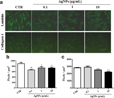
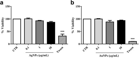
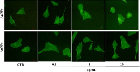
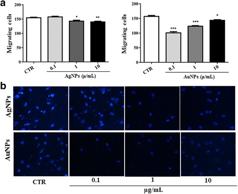
Nanoparticles have extremely wide applications in the medical and biological fields. They are being used in biosensors, local drug delivery, diagnostics, and medical therapy. However, the potential effects of nanoparticles on target cell and tissue function, apart from cytotoxicity, are not completely understood. Thus, the aim of this study was to investigate the in vitro effects of silver nanoparticles (AgNPs) and gold nanoparticles (AuNPs) on human fibroblasts with respect to their interaction with the extracellular matrix and in cell migration. Immunofluorescence analysis revealed that treatment with AgNPs or AuNPs decreased collagen and laminin production at all the concentrations tested (0.1, 1, and 10 μg/mL). Furthermore, cytofluorometric analysis showed that treatment with AgNPs reduced the percentage of cells expressing the collagen receptor very late antigen 2, αβ integrin (VLA-2) and the laminin receptor very late antigen 6, αβ integrin (VLA-6). In contrast, AuNP treatment increased and decreased the percentages of VLA-2-positive and VLA-6-positive cells, respectively, as compared to the findings for the controls. Analysis of cytoskeletal reorganization showed that treatment with both types of nanoparticles increased the formation of stress fibres and number of cell protrusions and impaired cell polarity. Fibroblasts exposed to different concentrations of AuNPs and AgNPs showed reduced migration through transwell chambers in the functional chemotaxis assay. These results demonstrated that metal nanoparticles may influence fibroblast function by negatively modulating the deposition of extracellular matrix molecules (ECM) and altering the expression of ECM receptors, cytoskeletal reorganization, and cell migration.
纳米颗粒在医学和生物领域有着极其广泛的应用。它们被用于生物传感器、局部药物递送、诊断和医学治疗。然而,除细胞毒性外,纳米颗粒对靶细胞和组织功能的潜在影响尚未完全明确。因此,本研究旨在探讨银纳米颗粒(AgNPs)和金纳米颗粒(AuNPs)在体外对人成纤维细胞与细胞外基质相互作用及细胞迁移方面的影响。免疫荧光分析显示,在所有测试浓度(0.1、1和10μg/mL)下,用AgNPs或AuNPs处理均会降低胶原蛋白和层粘连蛋白的产生。此外,细胞荧光分析表明,用AgNPs处理会降低表达胶原蛋白受体极晚期抗原2(αβ整合素,VLA - 2)和层粘连蛋白受体极晚期抗原6(αβ整合素,VLA - 6)的细胞百分比。相比之下,与对照组结果相比,AuNP处理分别增加和降低了VLA - 2阳性和VLA - 6阳性细胞的百分比。细胞骨架重组分析表明,两种纳米颗粒处理均会增加应力纤维的形成和细胞突起的数量,并损害细胞极性。在功能趋化性测定中,暴露于不同浓度AuNPs和AgNPs的成纤维细胞通过Transwell小室的迁移减少。这些结果表明,金属纳米颗粒可能通过负向调节细胞外基质分子(ECM)的沉积、改变ECM受体的表达、细胞骨架重组和细胞迁移来影响成纤维细胞功能。