Han Yang, Wang Shutao, Payen Thomas, Konofagou Elisa
Department of Biomedical Engineering, Columbia University, New York, NY, United States of America.
Phys Med Biol. 2017 Apr 21;62(8):3111-3123. doi: 10.1088/1361-6560/aa6024. Epub 2017 Mar 21.
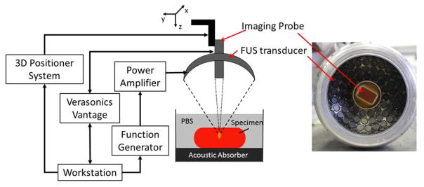
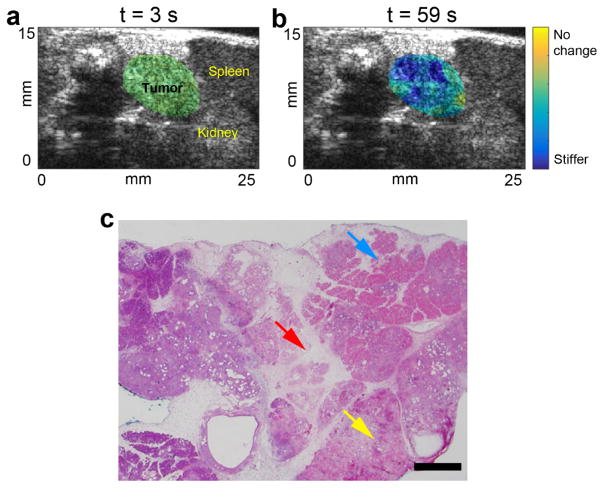
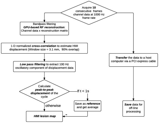
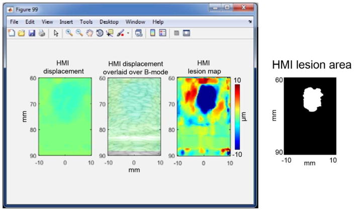
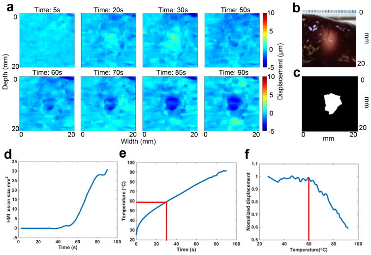
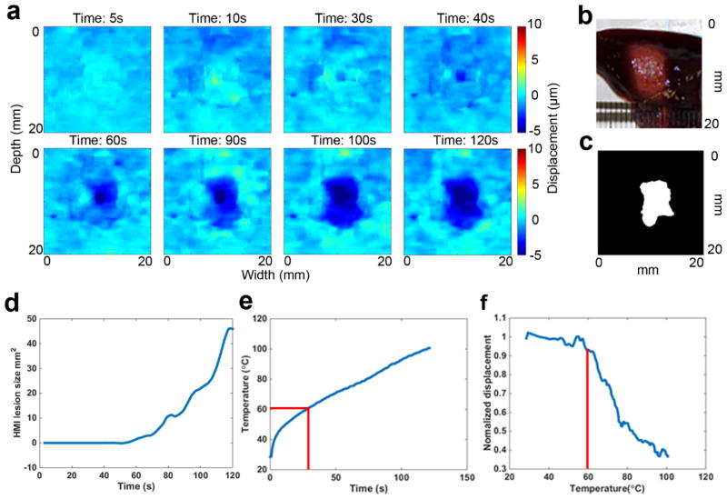
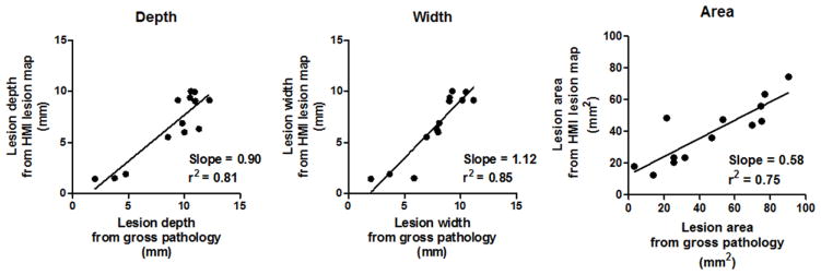
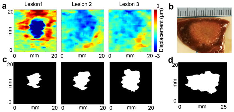
The successful clinical application of high intensity focused ultrasound (HIFU) ablation depends on reliable monitoring of the lesion formation. Harmonic motion imaging guided focused ultrasound (HMIgFUS) is an ultrasound-based elasticity imaging technique, which monitors HIFU ablation based on the stiffness change of the tissue instead of the echo intensity change in conventional B-mode monitoring, rendering it potentially more sensitive to lesion development. Our group has shown that predicting the lesion location based on the radiation force-excited region is feasible during HMIgFUS. In this study, the feasibility of a fast lesion mapping method is explored to directly monitor the lesion map during HIFU. The harmonic motion imaging (HMI) lesion map was generated by subtracting the reference HMI image from the present HMI peak-to-peak displacement map, as streamed on the computer display. The dimensions of the HMIgFUS lesions were compared against gross pathology. Excellent agreement was found between the lesion depth (r = 0.81, slope = 0.90), width (r = 0.85, slope = 1.12) and area (r = 0.58, slope = 0.75). In vivo feasibility was assessed in a mouse with a pancreatic tumor. These findings demonstrate that HMIgFUS can successfully map thermal lesions and monitor lesion development in real time in vitro and in vivo. The HMIgFUS technique may therefore constitute a novel clinical tool for HIFU treatment monitoring.
高强度聚焦超声(HIFU)消融术的成功临床应用依赖于对病灶形成的可靠监测。谐波运动成像引导聚焦超声(HMIgFUS)是一种基于超声的弹性成像技术,它基于组织硬度变化而非传统B模式监测中的回声强度变化来监测HIFU消融,这使其对病灶发展可能更敏感。我们团队已表明,在HMIgFUS过程中基于辐射力激发区域预测病灶位置是可行的。在本研究中,探索了一种快速病灶映射方法在HIFU期间直接监测病灶图的可行性。通过从计算机显示器上实时显示的当前HMI峰峰值位移图中减去参考HMI图像来生成谐波运动成像(HMI)病灶图。将HMIgFUS病灶的尺寸与大体病理学结果进行比较。发现病灶深度(r = 0.81,斜率 = 0.90)、宽度(r = 0.85,斜率 = 1.12)和面积(r = 0.58,斜率 = 0.75)之间具有极好的一致性。在一只患有胰腺肿瘤的小鼠中评估了体内可行性。这些发现表明,HMIgFUS能够成功绘制热损伤病灶图,并在体外和体内实时监测病灶发展。因此,HMIgFUS技术可能构成一种用于HIFU治疗监测的新型临床工具。