Gang Yadong, Zhou Hongfu, Jia Yao, Liu Ling, Liu Xiuli, Rao Gong, Li Longhui, Wang Xiaojun, Lv Xiaohua, Xiong Hanqing, Yang Zhongqin, Luo Qingming, Gong Hui, Zeng Shaoqun
Britton Chance Center for Biomedical Photonics, Wuhan National Laboratory for Optoelectronics-Huazhong University of Science and TechnologyWuhan, China; Key Laboratory of Biomedical Photonics of Ministry of Education, Department of Biomedical Engineering, Huazhong University of Science and TechnologyWuhan, China.
Front Neurosci. 2017 Mar 14;11:121. doi: 10.3389/fnins.2017.00121. eCollection 2017.
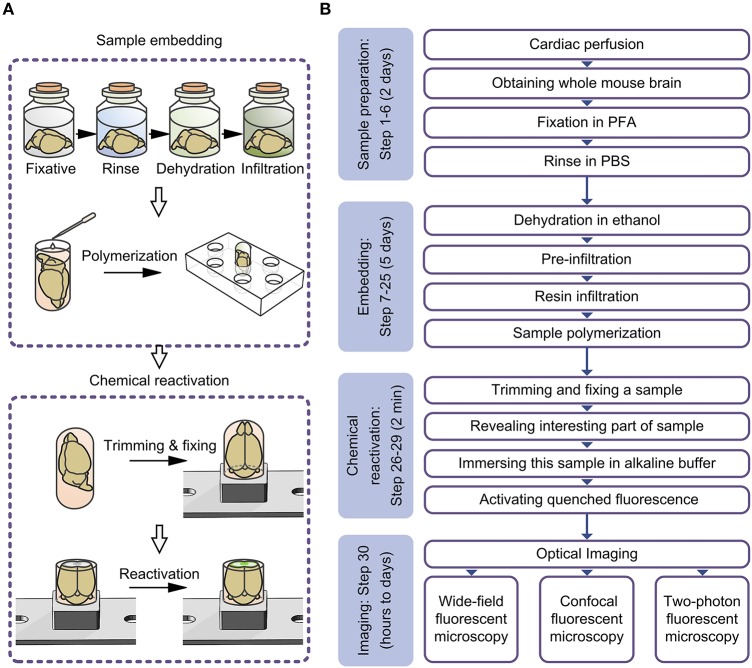
Resin embedding has been widely applied to fixing biological tissues for sectioning and imaging, but has long been regarded as incompatible with green fluorescent protein (GFP) labeled sample because it reduces fluorescence. Recently, it has been reported that resin-embedded GFP-labeled brain tissue can be imaged with high resolution. In this protocol, we describe an optimized protocol for resin embedding and chemical reactivation of fluorescent protein labeled mouse brain, we have used mice as experiment model, but the protocol should be applied to other species. This method involves whole brain embedding and chemical reactivation of the fluorescent signal in resin-embedded tissue. The whole brain embedding process takes a total of 7 days. The duration of chemical reactivation is ~2 min for penetrating 4 μm below the surface in the resin-embedded brain. This protocol provides an efficient way to prepare fluorescent protein labeled sample for high-resolution optical imaging. This kind of sample was demonstrated to be imaged by various optical micro-imaging methods. Fine structures labeled with GFP across a whole brain can be detected.
树脂包埋已被广泛应用于固定生物组织以进行切片和成像,但长期以来一直被认为与绿色荧光蛋白(GFP)标记的样本不相容,因为它会降低荧光。最近,有报道称树脂包埋的GFP标记脑组织可以进行高分辨率成像。在本方案中,我们描述了一种用于树脂包埋和荧光蛋白标记小鼠脑化学再激活的优化方案,我们以小鼠为实验模型,但该方案应适用于其他物种。该方法包括全脑包埋和树脂包埋组织中荧光信号的化学再激活。全脑包埋过程总共需要7天。在树脂包埋的大脑中,化学再激活持续约2分钟,以穿透表面以下4μm。本方案提供了一种为高分辨率光学成像制备荧光蛋白标记样本的有效方法。这种样本已被证明可以通过各种光学显微成像方法进行成像。可以检测到全脑范围内用GFP标记的精细结构。