Yanagiya Masahiro, Sato Masaaki, Kuwano Hideki, Nagayama Kazuhiro, Nakajima Jun
Department of Thoracic Surgery, University of Tokyo, Graduate School of Medicine, 7-3-1 Hongo, Bunkyo-ku, Tokyo, 113-8655, Japan.
Surg Case Rep. 2017 Dec;3(1):49. doi: 10.1186/s40792-017-0327-x. Epub 2017 Mar 28.
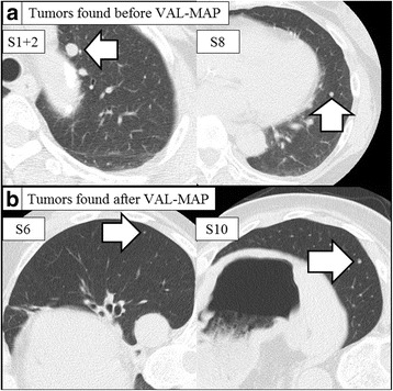
Virtual-assisted lung mapping is a novel bronchoscopic lung marking technique that uses virtual images to perform multiple concurrent dye marking of barely palpable pulmonary tumors. Subsequent chest computed tomography is required to confirm the locations marked. We here report a patient in whom computed tomography after virtual-assisted lung mapping unexpectedly revealed additional tiny pulmonary nodules.
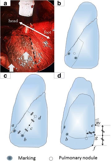
A 64-year-old woman with a history of renal cell carcinoma presented with two pulmonary nodules suspicious of metastases from renal cell carcinoma. Because we anticipated that the nodules would be difficult to palpate intraoperatively, we performed virtual-assisted lung mapping prior to attempting to resect them. Computed tomography after mapping unexpectedly detected two additional nodules. Although the existing markings did not relate to the newly found nodules, we used imaginary auxiliary lines and anatomical landmarks to extend the lung map to incorporate the unexpected nodules. The additional nodules were successfully resected by thoracoscopic wedge resection. Pathologic examination identified all nodules as metastases from renal cell carcinoma, and the surgical margins were negative.
Imaginary auxiliary lines and anatomical landmarks extended the existing lung map of virtual-assisted lung mapping, enabling resection of unexpected pulmonary nodules found in post-mapping computed tomography images.
虚拟辅助肺标测是一种新型的支气管镜肺标记技术,它利用虚拟图像对难以触及的肺部肿瘤进行多次同步染料标记。随后需要进行胸部计算机断层扫描以确认标记的位置。我们在此报告一例患者,在虚拟辅助肺标测后的计算机断层扫描意外发现了额外的微小肺结节。
一名64岁有肾细胞癌病史的女性患者出现两个可疑为肾细胞癌转移的肺结节。由于我们预计术中难以触及这些结节,因此在尝试切除之前进行了虚拟辅助肺标测。标测后的计算机断层扫描意外检测到另外两个结节。尽管现有的标记与新发现的结节无关,但我们使用假想辅助线和解剖标志将肺图扩展以纳入意外发现的结节。通过胸腔镜楔形切除术成功切除了额外的结节。病理检查确定所有结节均为肾细胞癌转移灶,手术切缘阴性。
假想辅助线和解剖标志扩展了虚拟辅助肺标测的现有肺图,使得能够切除在标测后计算机断层扫描图像中发现的意外肺结节。