Lei Lei, Tzekov Radouil, Li Huapeng, McDowell J Hugh, Gao Guangping, Smith W Clay, Tang Shibo, Kaushal Shalesh
State Key Laboratory of Ophthalmology, Zhongshan Ophthalmic Center, Sun Yat-sen University, No.54 South Xianlie Road, Guangzhou 510060, China.
Department of Ophthalmology, University of Massachusetts Medical School, 381 Plantation Street, Worcester, MA 01605, USA.
Int J Mol Sci. 2017 Mar 29;18(4):728. doi: 10.3390/ijms18040728.
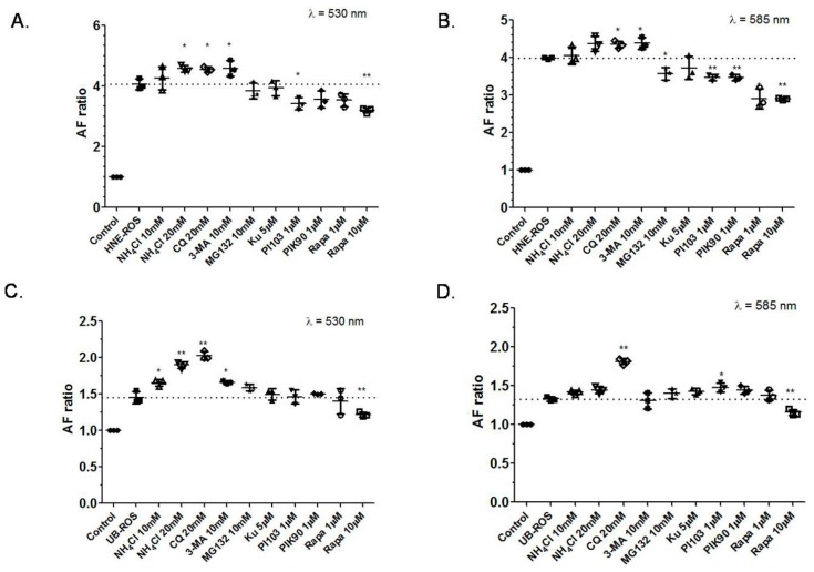
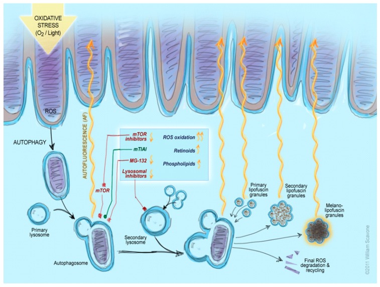
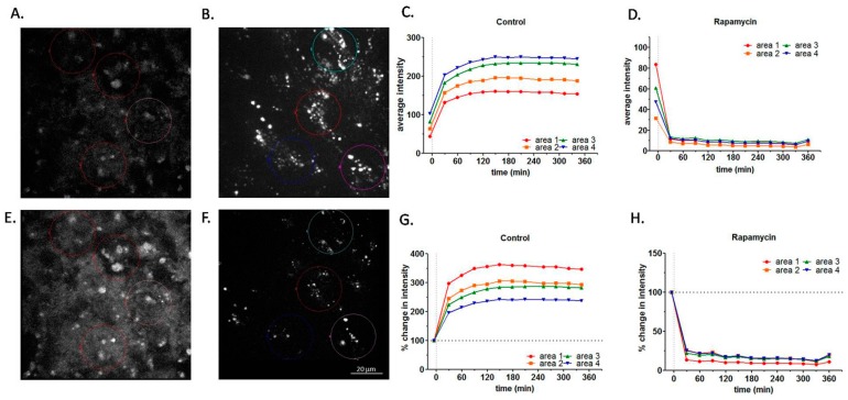
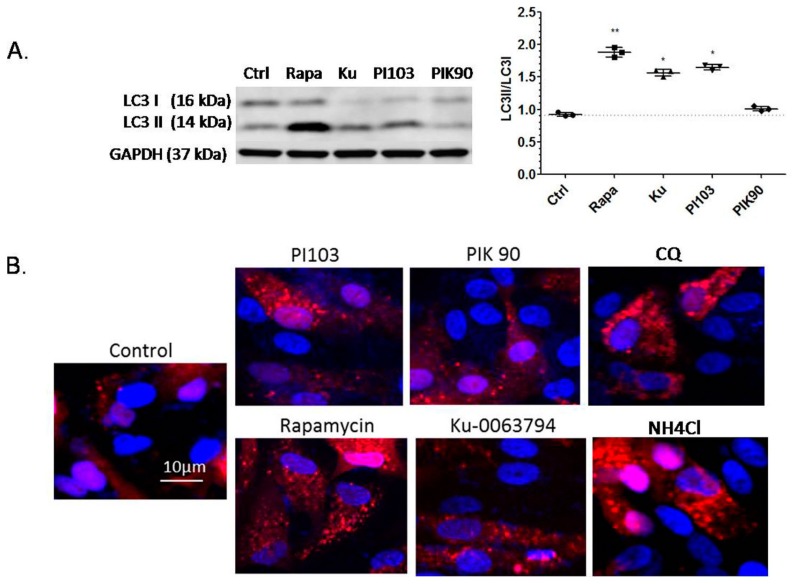
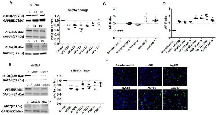
The accumulation of lipofuscin in the retinal pigment epithelium (RPE) is dependent on the effectiveness of photoreceptor outer segment material degradation. This study explored the role of autophagy in the fate of RPE lipofuscin degradation. After seven days of feeding with either native or modified rod outer segments, ARPE-19 cells were treated with enhancers or inhibitors of autophagy and the autofluorescence was detected by fluorescence-activated cell sorting. Supplementation with different types of rod outer segments increased lipofuscin-like autofluorescence (LLAF) after the inhibition of autophagy, while the induction of autophagy (e.g., application of rapamycin) decreased LLAF. The effects of autophagy induction were further confirmed by Western blotting, which showed the conversion of LC3-I to LC3-II, and by immunofluorescence microscopy, which detected the lysosomal activity of the autophagy inducers. We also monitored LLAF after the application of several autophagy inhibitors by RNA-interference and confocal microscopy. The results showed that, in general, the inhibition of the autophagy-related proteins resulted in an increase in LLAF when cells were fed with rod outer segments, which further confirms the effect of autophagy in the fate of RPE lipofuscin degradation. These results emphasize the complex role of autophagy in modulating RPE autofluorescence and confirm the possibility of the pharmacological clearance of RPE lipofuscin by small molecules.
视网膜色素上皮(RPE)中脂褐素的积累取决于光感受器外段物质降解的效率。本研究探讨了自噬在RPE脂褐素降解命运中的作用。在用天然或修饰的视杆外段喂养7天后,用自噬增强剂或抑制剂处理ARPE - 19细胞,并通过荧光激活细胞分选检测自发荧光。在抑制自噬后,补充不同类型的视杆外段会增加脂褐素样自发荧光(LLAF),而诱导自噬(如应用雷帕霉素)则会降低LLAF。通过蛋白质免疫印迹法(显示LC3 - I向LC3 - II的转化)和免疫荧光显微镜法(检测自噬诱导剂的溶酶体活性)进一步证实了自噬诱导的作用。我们还通过RNA干扰和共聚焦显微镜监测了几种自噬抑制剂应用后的LLAF。结果表明,总体而言,当细胞用视杆外段喂养时,抑制自噬相关蛋白会导致LLAF增加,这进一步证实了自噬在RPE脂褐素降解命运中的作用。这些结果强调了自噬在调节RPE自发荧光中的复杂作用,并证实了小分子药物清除RPE脂褐素的可能性。