• 文献检索
  • 文档翻译
  • 深度研究
  • 学术资讯
  • Suppr Zotero 插件Zotero 插件
  • 邀请有礼
  • 套餐&价格
  • 历史记录
应用&插件
Suppr Zotero 插件Zotero 插件浏览器插件Mac 客户端Windows 客户端微信小程序
定价
高级版会员购买积分包购买API积分包
服务
文献检索文档翻译深度研究API 文档MCP 服务
关于我们
关于 Suppr公司介绍联系我们用户协议隐私条款
关注我们

Suppr 超能文献

核心技术专利:CN118964589B侵权必究
粤ICP备2023148730 号-1Suppr @ 2026

文献检索

告别复杂PubMed语法,用中文像聊天一样搜索,搜遍4000万医学文献。AI智能推荐,让科研检索更轻松。

立即免费搜索

文件翻译

保留排版,准确专业,支持PDF/Word/PPT等文件格式,支持 12+语言互译。

免费翻译文档

深度研究

AI帮你快速写综述,25分钟生成高质量综述,智能提取关键信息,辅助科研写作。

立即免费体验

量化色素性视网膜炎患者的眼底自发荧光

Quantifying Fundus Autofluorescence in Patients With Retinitis Pigmentosa.

作者信息

Schuerch Kaspar, Woods Russell L, Lee Winston, Duncker Tobias, Delori François C, Allikmets Rando, Tsang Stephen H, Sparrow Janet R

机构信息

Department of Ophthalmology, Columbia University, New York, New York, United States.

Schepens Eye Research Institute and Department of Ophthalmology, Harvard Medical School, Boston, Massachusetts, United States.

出版信息

Invest Ophthalmol Vis Sci. 2017 Mar 1;58(3):1843-1855. doi: 10.1167/iovs.16-21302.

DOI:10.1167/iovs.16-21302
PMID:28358950
原文链接:https://pmc.ncbi.nlm.nih.gov/articles/PMC5377994/
Abstract

PURPOSE

Using quantitative fundus autofluorescence (qAF), we analyzed short-wavelength autofluorescent (SW-AF) rings in RP.

METHODS

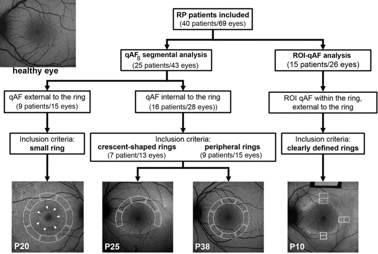
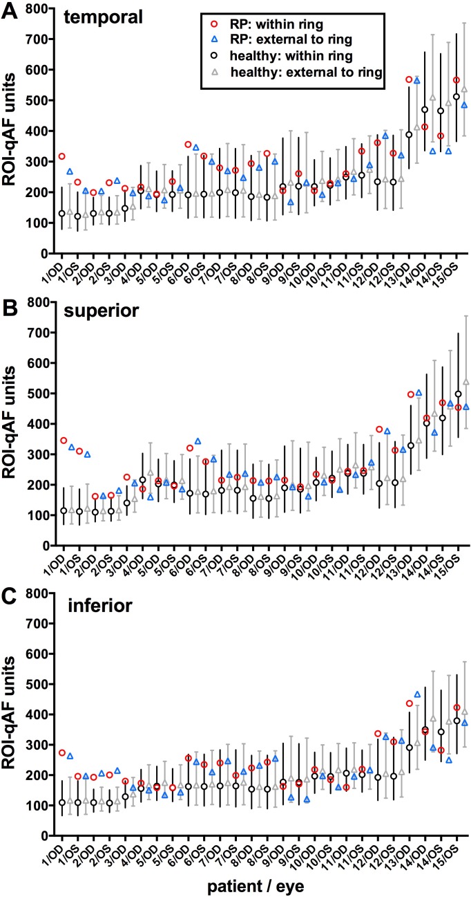
Short-wavelength autofluorescent images (486 nm excitation) of 40 patients with RP (69 eyes) were acquired with a confocal scanning laser ophthalmoscope equipped with an internal fluorescent reference. Mean qAF was measured in eight preset segments (qAF8) and in region of interest (ROI)-qAF (200-700 μm) within and external to the borders of the rings at superior, temporal, and inferior sites relative to the ring. For both groups, qAF in patients with RP was compared to age-similar and race/ethnicity-matched healthy eyes at equivalent retinal locations.

RESULTS

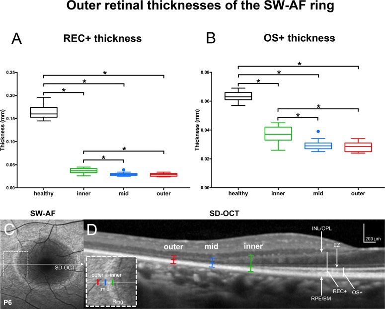
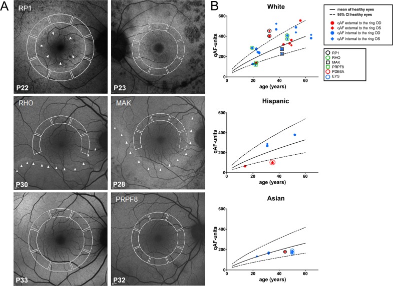
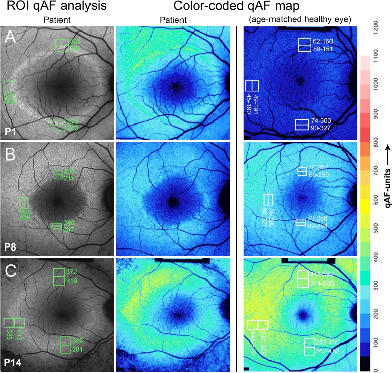
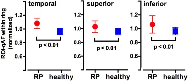
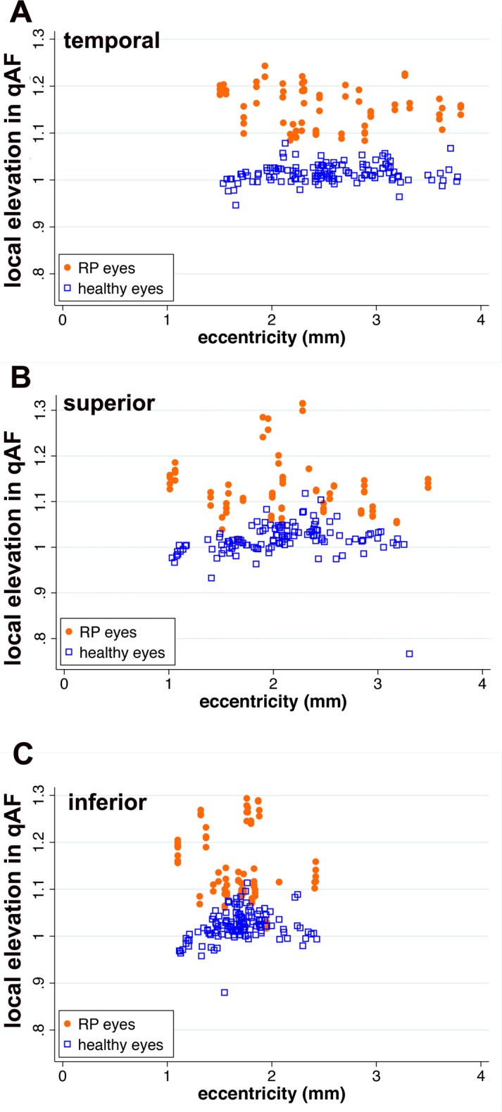
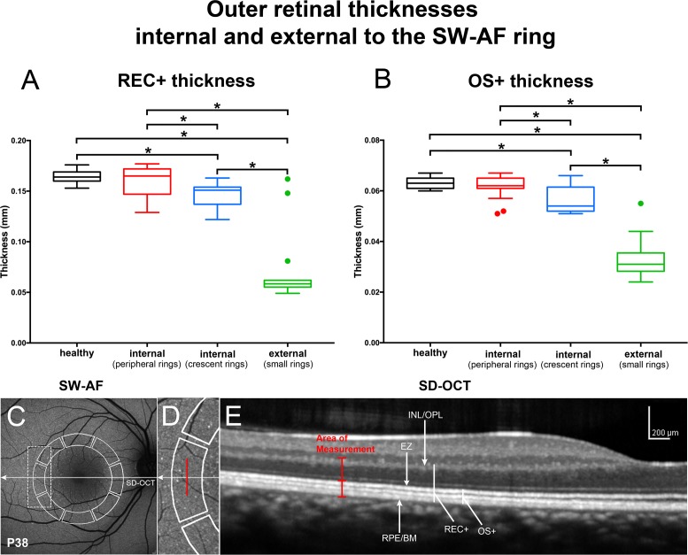
In 71% of eyes of RP patients, qAF8 acquired internal to the inner border of the ring, was within the 95% confidence interval (CI) for healthy eyes, while in the remaining RP eyes qAF8 was either higher or lower than the CI. Measured external to the ring, qAF8 values were within the CI in 47% of RP eyes with the other eyes being higher or lower. In 28% of sites measured by ROI-qAF within the SW-AF ring, values were above the 95% CI of healthy controls. Region of interest-qAF measured just external to the ring was within the CI of healthy eyes in 74% of locations. The average local elevation in qAF within the ring was approximately 15%. In SD-OCT scans, photoreceptor-attributable reflectivity bands were thinned within and external to the ring.

CONCLUSIONS

Increased fluorophore production may be a factor in the formation of the SW-AF rings in RP.

摘要

目的

我们使用定量眼底自发荧光(qAF)分析了视网膜色素变性(RP)中的短波自发荧光(SW-AF)环。

方法

使用配备内部荧光参考的共焦扫描激光检眼镜获取40例RP患者(69只眼)的短波自发荧光图像(486nm激发)。在相对于环的上方、颞侧和下方部位,在环边界内和外的八个预设节段(qAF8)以及感兴趣区域(ROI)-qAF(200-700μm)中测量平均qAF。对于两组,将RP患者的qAF与年龄相似且种族/民族匹配的健康眼在等效视网膜位置进行比较。

结果

在RP患者71%的眼中,在环内边界内部获取的qAF8在健康眼的95%置信区间(CI)内,而在其余的RP眼中,qAF8要么高于要么低于CI。在环外测量时,47%的RP眼的qAF8值在CI内,其他眼则更高或更低。在SW-AF环内通过ROI-qAF测量的28%的部位,值高于健康对照的95%CI。在环外紧邻处测量的感兴趣区域-qAF在74%的位置在健康眼的CI内。环内qAF的平均局部升高约为15%。在SD-OCT扫描中,环内和环外可归因于光感受器的反射带变薄。

结论

荧光团产生增加可能是RP中SW-AF环形成的一个因素。

https://cdn.ncbi.nlm.nih.gov/pmc/blobs/095a/5377994/cf207586e094/i1552-5783-58-3-1843-f09.jpg
https://cdn.ncbi.nlm.nih.gov/pmc/blobs/095a/5377994/ed8507f537ff/i1552-5783-58-3-1843-f01.jpg
https://cdn.ncbi.nlm.nih.gov/pmc/blobs/095a/5377994/543243c1ae4a/i1552-5783-58-3-1843-f02.jpg
https://cdn.ncbi.nlm.nih.gov/pmc/blobs/095a/5377994/51cfa62d9d53/i1552-5783-58-3-1843-f03.jpg
https://cdn.ncbi.nlm.nih.gov/pmc/blobs/095a/5377994/d2194728c814/i1552-5783-58-3-1843-f04.jpg
https://cdn.ncbi.nlm.nih.gov/pmc/blobs/095a/5377994/17ecb69b2428/i1552-5783-58-3-1843-f05.jpg
https://cdn.ncbi.nlm.nih.gov/pmc/blobs/095a/5377994/5faa07a3be06/i1552-5783-58-3-1843-f06.jpg
https://cdn.ncbi.nlm.nih.gov/pmc/blobs/095a/5377994/fc86628d9530/i1552-5783-58-3-1843-f07.jpg
https://cdn.ncbi.nlm.nih.gov/pmc/blobs/095a/5377994/0c1ee2d23394/i1552-5783-58-3-1843-f08.jpg
https://cdn.ncbi.nlm.nih.gov/pmc/blobs/095a/5377994/cf207586e094/i1552-5783-58-3-1843-f09.jpg

相似文献

1
Quantifying Fundus Autofluorescence in Patients With Retinitis Pigmentosa.量化色素性视网膜炎患者的眼底自发荧光
Invest Ophthalmol Vis Sci. 2017 Mar 1;58(3):1843-1855. doi: 10.1167/iovs.16-21302.
2
Quantitative Fundus Autofluorescence and Genetic Associations in Macular, Cone, and Cone-Rod Dystrophies.定量眼底自发荧光与黄斑、视锥和视锥-杆营养不良的遗传相关性。
Ophthalmol Retina. 2020 Jul;4(7):737-749. doi: 10.1016/j.oret.2020.02.009. Epub 2020 Feb 27.
3
Quantitative Fundus Autofluorescence in Pseudoxanthoma Elasticum.弹性假黄瘤的定量眼底自发荧光
Invest Ophthalmol Vis Sci. 2017 Dec 1;58(14):6159-6165. doi: 10.1167/iovs.17-22007.
4
Repeatability and reliability of quantitative fundus autofluorescence imaging in patients with early and intermediate age-related macular degeneration.定量眼底自发荧光成像在早期和中期年龄相关性黄斑变性患者中的重复性和可靠性。
Acta Ophthalmol. 2019 Jun;97(4):e526-e532. doi: 10.1111/aos.13987. Epub 2018 Dec 13.
5
Quantitative Fundus Autofluorescence in Rhesus Macaques in Aging and Age-Related Drusen.定量眼底自发荧光在衰老和年龄相关性玻璃膜疣的恒河猴模型中的应用。
Invest Ophthalmol Vis Sci. 2020 Jul 1;61(8):16. doi: 10.1167/iovs.61.8.16.
6
Central Serous Chorioretinopathy Analyzed by Multimodal Imaging.多模态成像分析中心性浆液性脉络膜视网膜病变。
Transl Vis Sci Technol. 2021 Jan 8;10(1):15. doi: 10.1167/tvst.10.1.15. eCollection 2021 Jan.
7
Quantitative fundus autofluorescence and optical coherence tomography in best vitelliform macular dystrophy.定量眼底自发荧光和光学相干断层扫描在最佳型类玻璃体黄斑营养不良中的应用。
Invest Ophthalmol Vis Sci. 2014 Mar 13;55(3):1471-82. doi: 10.1167/iovs.13-13834.
8
Quantitative Fundus Autofluorescence and Optical Coherence Tomography in ABCA4 Carriers.ABCA4基因携带者的定量眼底自发荧光和光学相干断层扫描
Invest Ophthalmol Vis Sci. 2015 Nov;56(12):7274-85. doi: 10.1167/iovs.15-17371.
9
QUANTITATIVE AUTOFLUORESCENCE IN CENTRAL SEROUS CHORIORETINOPATHY.中心性浆液性脉络膜视网膜病变的定量自发荧光。
Retina. 2024 May 1;44(5):844-851. doi: 10.1097/IAE.0000000000004029.
10
Quantitative fundus autofluorescence in recessive Stargardt disease.隐性Stargardt病的定量眼底自发荧光
Invest Ophthalmol Vis Sci. 2014 May 1;55(5):2841-52. doi: 10.1167/iovs.13-13624.

引用本文的文献

1
Bisretinoid lipofuscin, fundus autofluorescence and retinal disease.双视黄醛脂褐质、眼底自发荧光与视网膜疾病。
Prog Retin Eye Res. 2025 Jul 8;108:101388. doi: 10.1016/j.preteyeres.2025.101388.
2
Characterization of Macular Fundus Autofluorescence Changes in Patients with Retinitis Pigmentosa.视网膜色素变性患者黄斑区眼底自发荧光变化的特征分析
Ophthalmic Res. 2025;68(1):156-168. doi: 10.1159/000543082. Epub 2025 Jan 31.
3
Fluorescence Lifetime Imaging of Human Retinal Pigment Epithelium in Pentosan Polysulfate Toxicity Using Adaptive Optics Scanning Light Ophthalmoscopy.

本文引用的文献

1
MULTIMODAL IMAGING OF DISEASE-ASSOCIATED PIGMENTARY CHANGES IN RETINITIS PIGMENTOSA.色素性视网膜炎中疾病相关色素变化的多模态成像
Retina. 2016 Dec;36 Suppl 1(Suppl 1):S147-S158. doi: 10.1097/IAE.0000000000001256.
2
Photodegradation of retinal bisretinoids in mouse models and implications for macular degeneration.小鼠模型中视网膜双视黄醛的光降解及其对黄斑变性的影响。
Proc Natl Acad Sci U S A. 2016 Jun 21;113(25):6904-9. doi: 10.1073/pnas.1524774113. Epub 2016 Jun 6.
3
Presence of a Triple Concentric Autofluorescence Ring in NR2E3-p.G56R-Linked Autosomal Dominant Retinitis Pigmentosa (ADRP).
自适应光学扫描激光检眼镜观察戊聚糖多硫酸盐毒性对人视网膜色素上皮细胞的荧光寿命成像。
Invest Ophthalmol Vis Sci. 2024 Apr 1;65(4):27. doi: 10.1167/iovs.65.4.27.
4
Lipofuscin, Its Origin, Properties, and Contribution to Retinal Fluorescence as a Potential Biomarker of Oxidative Damage to the Retina.脂褐素,其起源、特性以及作为视网膜氧化损伤潜在生物标志物对视网膜荧光的贡献。
Antioxidants (Basel). 2023 Dec 13;12(12):2111. doi: 10.3390/antiox12122111.
5
Primary versus Secondary Elevations in Fundus Autofluorescence.眼底自发荧光的原发性与继发性抬高。
Int J Mol Sci. 2023 Aug 2;24(15):12327. doi: 10.3390/ijms241512327.
6
New insights in the multimodal imaging of retinitis pigmentosa.色素性视网膜炎的多模态成像新见解。
Eur J Ophthalmol. 2024 Mar;34(2):357-366. doi: 10.1177/11206721231172863. Epub 2023 Apr 27.
7
Morphological and functional involvement of the inner retina in retinitis pigmentosa.色素性视网膜炎中内视网膜的形态和功能改变。
Eye (Lond). 2023 May;37(7):1424-1431. doi: 10.1038/s41433-022-02139-7. Epub 2022 Jun 29.
8
Genetic, environmental and other risk factors for progression of retinitis pigmentosa.视网膜色素变性进展的遗传、环境及其他风险因素。
Int J Ophthalmol. 2022 May 18;15(5):828-837. doi: 10.18240/ijo.2022.05.21. eCollection 2022.
9
Quantitative autofluorescence findings in patients undergoing hydroxychloroquine treatment.接受羟氯喹治疗的患者的定量自发荧光研究结果。
Clin Exp Ophthalmol. 2022 Jul;50(5):500-509. doi: 10.1111/ceo.14090. Epub 2022 May 14.
10
Long-term vitamin A supplementation in a preclinical mouse model for RhoD190N-associated retinitis pigmentosa.RhoD190N 相关性视网膜色素变性的临床前小鼠模型中长期补充维生素 A。
Hum Mol Genet. 2022 Jul 21;31(14):2438-2451. doi: 10.1093/hmg/ddac032.
NR2E3-p.G56R相关常染色体显性遗传性视网膜色素变性(ADRP)中三重同心自发荧光环的存在
Invest Ophthalmol Vis Sci. 2016 Apr 1;57(4):2001-2. doi: 10.1167/iovs.16-19459.
4
Quantitative Fundus Autofluorescence and Optical Coherence Tomography in ABCA4 Carriers.ABCA4基因携带者的定量眼底自发荧光和光学相干断层扫描
Invest Ophthalmol Vis Sci. 2015 Nov;56(12):7274-85. doi: 10.1167/iovs.15-17371.
5
Flecks in Recessive Stargardt Disease: Short-Wavelength Autofluorescence, Near-Infrared Autofluorescence, and Optical Coherence Tomography.隐性Stargardt病中的斑点:短波自发荧光、近红外自发荧光和光学相干断层扫描
Invest Ophthalmol Vis Sci. 2015 Jul;56(8):5029-39. doi: 10.1167/iovs.15-16763.
6
Multimodal Imaging of Central Retinal Disease Progression in a 2-Year Mean Follow-up of Retinitis Pigmentosa.色素性视网膜炎2年平均随访中视网膜中央疾病进展的多模态成像
Am J Ophthalmol. 2015 Oct;160(4):786-98.e4. doi: 10.1016/j.ajo.2015.06.032. Epub 2015 Jul 9.
7
Quantitative Fundus Autofluorescence and Optical Coherence Tomography in PRPH2/RDS- and ABCA4-Associated Disease Exhibiting Phenotypic Overlap.在表现出表型重叠的PRPH2/RDS和ABCA4相关疾病中定量眼底自发荧光和光学相干断层扫描
Invest Ophthalmol Vis Sci. 2015 May;56(5):3159-70. doi: 10.1167/iovs.14-16343.
8
Rates of decline in regions of the visual field defined by frequency-domain optical coherence tomography in patients with RPGR-mediated X-linked retinitis pigmentosa.在RPGR介导的X连锁视网膜色素变性患者中,通过频域光学相干断层扫描定义的视野区域的下降率。
Ophthalmology. 2015 Apr;122(4):833-9. doi: 10.1016/j.ophtha.2014.11.005. Epub 2014 Dec 31.
9
ISCEV Standard for full-field clinical electroretinography (2015 update).国际临床视觉电生理学会全视野临床视网膜电图标准(2015年更新版)
Doc Ophthalmol. 2015 Feb;130(1):1-12. doi: 10.1007/s10633-014-9473-7. Epub 2014 Dec 14.
10
A comparison of progressive loss of the ellipsoid zone (EZ) band in autosomal dominant and x-linked retinitis pigmentosa.常染色体显性遗传性视网膜色素变性与X连锁视网膜色素变性中椭圆体带(EZ)逐渐丧失的比较
Invest Ophthalmol Vis Sci. 2014 Oct 23;55(11):7417-22. doi: 10.1167/iovs.14-15013.