• 文献检索
  • 文档翻译
  • 深度研究
  • 学术资讯
  • Suppr Zotero 插件Zotero 插件
  • 邀请有礼
  • 套餐&价格
  • 历史记录
应用&插件
Suppr Zotero 插件Zotero 插件浏览器插件Mac 客户端Windows 客户端微信小程序
定价
高级版会员购买积分包购买API积分包
服务
文献检索文档翻译深度研究API 文档MCP 服务
关于我们
关于 Suppr公司介绍联系我们用户协议隐私条款
关注我们

Suppr 超能文献

核心技术专利:CN118964589B侵权必究
粤ICP备2023148730 号-1Suppr @ 2026

文献检索

告别复杂PubMed语法,用中文像聊天一样搜索,搜遍4000万医学文献。AI智能推荐,让科研检索更轻松。

立即免费搜索

文件翻译

保留排版,准确专业,支持PDF/Word/PPT等文件格式,支持 12+语言互译。

免费翻译文档

深度研究

AI帮你快速写综述,25分钟生成高质量综述,智能提取关键信息,辅助科研写作。

立即免费体验

基于血管特征的 3D 血管周围空间分割的结构化学习。

Structured Learning for 3-D Perivascular Space Segmentation Using Vascular Features.

出版信息

IEEE Trans Biomed Eng. 2017 Dec;64(12):2803-2812. doi: 10.1109/TBME.2016.2638918. Epub 2017 Mar 1.

DOI:10.1109/TBME.2016.2638918
PMID:28362579
原文链接:https://pmc.ncbi.nlm.nih.gov/articles/PMC5749233/
Abstract

OBJECTIVE

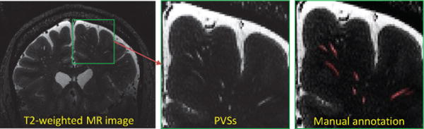
The goal of this paper is to automatically segment perivascular spaces (PVSs) in brain from high-resolution 7T magnetic resonance (MR) images.

METHODS

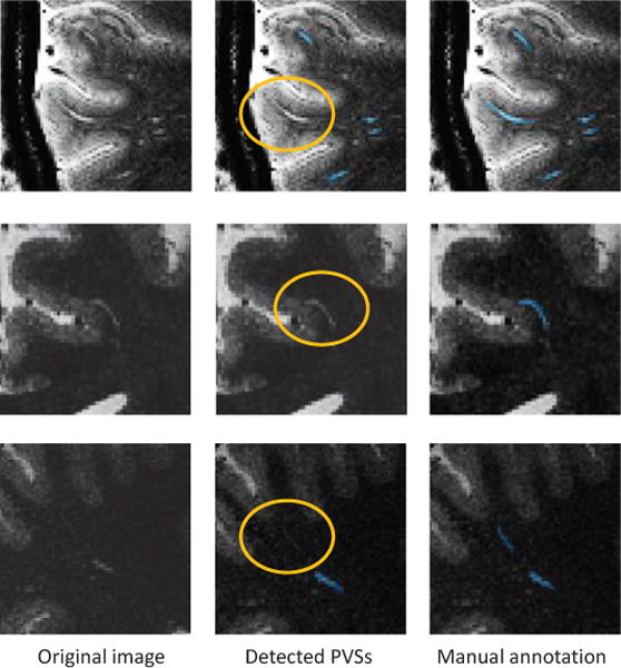
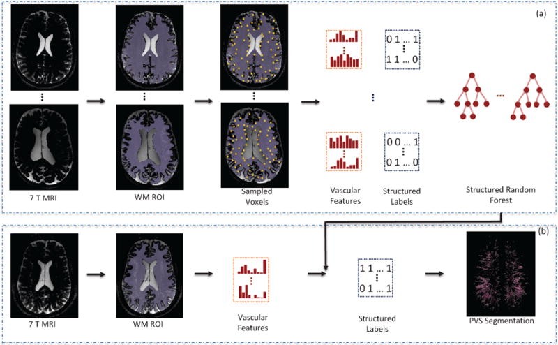
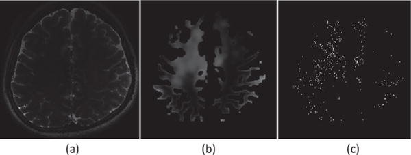
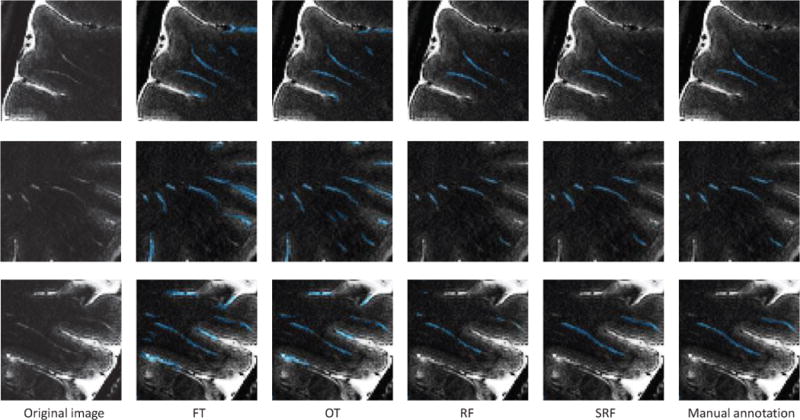
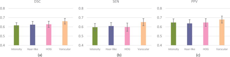
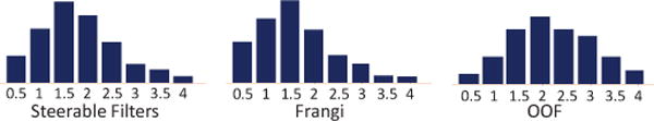
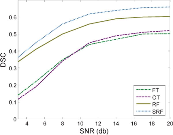
We propose a structured-learning-based segmentation framework to extract the PVSs from high-resolution 7T MR images. Specifically, we integrate three types of vascular filter responses into a structured random forest for classifying voxels into two categories, i.e., PVS and background. In addition, we propose a novel entropy-based sampling strategy to extract informative samples in the background for training an explicit classification model. Since the vascular filters can extract various vascular features, even thin and low-contrast structures can be effectively extracted from noisy backgrounds. Moreover, continuous and smooth segmentation results can be obtained by utilizing patch-based structured labels.

RESULTS

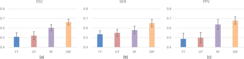
The performance of our proposed method is evaluated on 19 subjects with 7T MR images, with the Dice similarity coefficient reaching 66%.

CONCLUSION

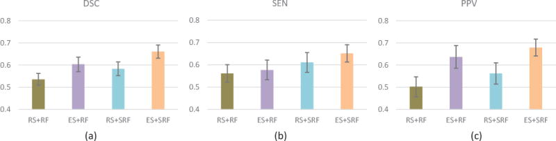
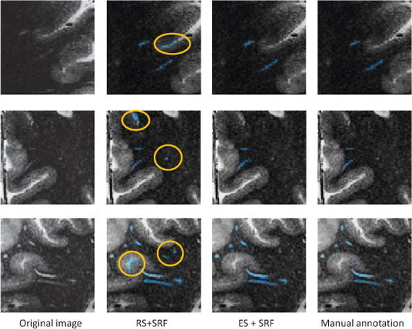
The joint use of entropy-based sampling strategy, vascular features, and structured learning can improve the segmentation accuracy.

SIGNIFICANCE

Instead of manual annotation, our method provides an automatic way for PVS segmentation. Moreover, our method can be potentially used for other vascular structure segmentation because of its data-driven property.

摘要

目的

本文旨在从高分辨率 7T 磁共振(MR)图像中自动分割脑内血管周围间隙(PVS)。

方法

我们提出了一种基于结构学习的分割框架,从高分辨率 7T MR 图像中提取 PVS。具体来说,我们将三种类型的血管滤波器响应集成到一个结构随机森林中,将体素分类为 PVS 和背景两类。此外,我们提出了一种新的基于熵的采样策略,从背景中提取信息丰富的样本,以训练显式分类模型。由于血管滤波器可以提取各种血管特征,即使是薄的和低对比度的结构也可以从噪声背景中有效提取。此外,通过利用基于补丁的结构标签,可以获得连续平滑的分割结果。

结果

我们在 19 名受试者的 7T MR 图像上评估了所提出方法的性能,Dice 相似系数达到 66%。

结论

基于熵的采样策略、血管特征和结构学习的联合使用可以提高分割准确性。

意义

与手动标注相比,我们的方法为 PVS 分割提供了一种自动的方法。此外,由于其数据驱动的特性,我们的方法可以潜在地用于其他血管结构的分割。

https://cdn.ncbi.nlm.nih.gov/pmc/blobs/cffa/5749233/b598cec946c7/nihms923369f11.jpg
https://cdn.ncbi.nlm.nih.gov/pmc/blobs/cffa/5749233/84bc3e2c3cca/nihms923369f1.jpg
https://cdn.ncbi.nlm.nih.gov/pmc/blobs/cffa/5749233/9cfe689d9db9/nihms923369f2.jpg
https://cdn.ncbi.nlm.nih.gov/pmc/blobs/cffa/5749233/ca6fb6668bd5/nihms923369f3.jpg
https://cdn.ncbi.nlm.nih.gov/pmc/blobs/cffa/5749233/6d5dfd4c7813/nihms923369f4.jpg
https://cdn.ncbi.nlm.nih.gov/pmc/blobs/cffa/5749233/b87ec07d7cc2/nihms923369f5.jpg
https://cdn.ncbi.nlm.nih.gov/pmc/blobs/cffa/5749233/0259ec37d953/nihms923369f6.jpg
https://cdn.ncbi.nlm.nih.gov/pmc/blobs/cffa/5749233/3fde6b78f92b/nihms923369f7.jpg
https://cdn.ncbi.nlm.nih.gov/pmc/blobs/cffa/5749233/8e8a18085250/nihms923369f8.jpg
https://cdn.ncbi.nlm.nih.gov/pmc/blobs/cffa/5749233/57aacd076198/nihms923369f9.jpg
https://cdn.ncbi.nlm.nih.gov/pmc/blobs/cffa/5749233/d5f678d67206/nihms923369f10.jpg
https://cdn.ncbi.nlm.nih.gov/pmc/blobs/cffa/5749233/b598cec946c7/nihms923369f11.jpg

相似文献

1
Structured Learning for 3-D Perivascular Space Segmentation Using Vascular Features.基于血管特征的 3D 血管周围空间分割的结构化学习。
IEEE Trans Biomed Eng. 2017 Dec;64(12):2803-2812. doi: 10.1109/TBME.2016.2638918. Epub 2017 Mar 1.
2
Segmentation of Perivascular Spaces Using Vascular Features and Structured Random Forest from 7T MR Image.基于7T磁共振图像利用血管特征和结构化随机森林分割血管周围间隙
Mach Learn Med Imaging. 2016 Oct;10019:61-68. doi: 10.1007/978-3-319-47157-0_8. Epub 2016 Oct 1.
3
Multi-channel multi-scale fully convolutional network for 3D perivascular spaces segmentation in 7T MR images.多通道多尺度全卷积网络用于 7T MR 图像中血管周围空间的分割。
Med Image Anal. 2018 May;46:106-117. doi: 10.1016/j.media.2018.02.009. Epub 2018 Feb 27.
4
Segmentation of perivascular spaces in 7T MR image using auto-context model with orientation-normalized features.使用具有方向归一化特征的自动上下文模型对7T磁共振图像中的血管周围间隙进行分割。
Neuroimage. 2016 Jul 1;134:223-235. doi: 10.1016/j.neuroimage.2016.03.076. Epub 2016 Apr 1.
5
Learning-based 3T brain MRI segmentation with guidance from 7T MRI labeling.基于学习的3T脑磁共振成像分割,由7T磁共振成像标记引导。
Med Phys. 2016 Dec;43(12):6588-6597. doi: 10.1118/1.4967487.
6
Learning-based 3T brain MRI segmentation with guidance from 7T MRI labeling.基于学习的3T脑磁共振成像分割,并以7T磁共振成像标记为指导。
Med Phys. 2016 Dec;43(12):6588. doi: 10.1118/1.4967487.
7
The feasibility of quantitative MRI of perivascular spaces at 7T.7T下血管周围间隙定量磁共振成像的可行性
J Neurosci Methods. 2015 Dec 30;256:151-6. doi: 10.1016/j.jneumeth.2015.09.001. Epub 2015 Sep 8.
8
The Learning-based Automatic Segmentation Algorithm of Brain MR Images Based on 7T.基于 7T 的脑磁共振图像的基于学习的自动分割算法。
Curr Med Imaging. 2021;17(3):342-351. doi: 10.2174/1573405616666200806171509.
9
Deep-learning-based segmentation of perivascular spaces on T2-Weighted 3T magnetic resonance images.基于深度学习的3T加权T2磁共振图像上血管周围间隙的分割
Front Aging Neurosci. 2024 Aug 29;16:1457405. doi: 10.3389/fnagi.2024.1457405. eCollection 2024.
10
Deformable segmentation of 3D MR prostate images via distributed discriminative dictionary and ensemble learning.通过分布式判别字典和集成学习实现3D磁共振前列腺图像的可变形分割
Med Phys. 2014 Jul;41(7):072303. doi: 10.1118/1.4884224.

引用本文的文献

1
Beyond the cerebellum: perivascular space burden in spinocerebellar ataxia type 3 extends to multiple brain regions.小脑之外:3型脊髓小脑共济失调的血管周围间隙病变累及多个脑区。
Brain Commun. 2025 Mar 27;7(2):fcaf118. doi: 10.1093/braincomms/fcaf118. eCollection 2025.
2
Perivascular space imaging during therapy for medulloblastoma.髓母细胞瘤治疗期间的血管周围间隙成像
PLoS One. 2025 Feb 7;20(2):e0318278. doi: 10.1371/journal.pone.0318278. eCollection 2025.
3
Deep-learning-based segmentation of perivascular spaces on T2-Weighted 3T magnetic resonance images.

本文引用的文献

1
Assessment of hessian-based Frangi vesselness filter in optoacoustic imaging.基于hessian的Frangi血管造影滤波器在光声成像中的评估。
Photoacoustics. 2020 Jul 13;20:100200. doi: 10.1016/j.pacs.2020.100200. eCollection 2020 Dec.
2
Pulmonary Fissure Detection in CT Images Using a Derivative of Stick Filter.基于 stick 滤波器导数的 CT 图像肺裂检测
IEEE Trans Med Imaging. 2016 Jun;35(6):1488-500. doi: 10.1109/TMI.2016.2517680. Epub 2016 Jan 13.
3
Visualization of perivascular spaces in the human brain at 7T: sequence optimization and morphology characterization.
基于深度学习的3T加权T2磁共振图像上血管周围间隙的分割
Front Aging Neurosci. 2024 Aug 29;16:1457405. doi: 10.3389/fnagi.2024.1457405. eCollection 2024.
4
Quantitative assessment of enlarged perivascular spaces via deep-learning in community-based older adults reveals independent associations with vascular neuropathologies, vascular risk factors and cognition.通过深度学习对社区老年人群中扩大的血管周围间隙进行定量评估,揭示了其与血管神经病理学、血管危险因素和认知之间的独立关联。
Brain Commun. 2024 Jul 30;6(4):fcae252. doi: 10.1093/braincomms/fcae252. eCollection 2024.
5
Overview of the Current Knowledge and Conventional MRI Characteristics of Peri- and Para-Vascular Spaces.血管周围间隙和血管旁间隙的当前知识概述及传统MRI特征
Brain Sci. 2024 Jan 28;14(2):138. doi: 10.3390/brainsci14020138.
6
Perivascular spaces relate to the course and cognition of Huntington's disease.血管周围间隙与亨廷顿舞蹈病的病程及认知情况相关。
Transl Neurodegener. 2023 May 15;12(1):30. doi: 10.1186/s40035-023-00359-9.
7
Perivascular network segmentations derived from high-field MRI and their implications for perivascular and parenchymal mass transport in the rat brain.从高场 MRI 得出的血管周围网络分割及其对大鼠脑血管周围和实质物质转运的影响。
Sci Rep. 2023 Jun 6;13(1):9205. doi: 10.1038/s41598-023-34850-0.
8
Deep Learning Based Detection of Enlarged Perivascular Spaces on Brain MRI.基于深度学习的脑磁共振成像上血管周围间隙增宽的检测
Neuroimage Rep. 2023 Mar;3(1). doi: 10.1016/j.ynirp.2023.100162. Epub 2023 Mar 7.
9
Characteristics of perivascular space dilatation in normal aging.正常衰老过程中血管周围间隙扩张的特征。
Hum Brain Mapp. 2023 Jun 1;44(8):3232-3240. doi: 10.1002/hbm.26277. Epub 2023 Mar 17.
10
Weakly supervised perivascular spaces segmentation with salient guidance of Frangi filter.基于 Frangi 滤波器显著引导的弱监督血管周围空间分割。
Magn Reson Med. 2023 Jun;89(6):2419-2431. doi: 10.1002/mrm.29593. Epub 2023 Jan 24.
7T下人脑血管周围间隙的可视化:序列优化与形态学特征分析
Neuroimage. 2016 Jan 15;125:895-902. doi: 10.1016/j.neuroimage.2015.10.078. Epub 2015 Oct 28.
4
Virchow-Robin Spaces: Correlations with Polysomnography-Derived Sleep Parameters.魏尔啸-罗宾间隙:与多导睡眠图衍生的睡眠参数的相关性
Sleep. 2015 Jun 1;38(6):853-8. doi: 10.5665/sleep.4726.
5
Iterative Vessel Segmentation of Fundus Images.眼底图像的迭代血管分割
IEEE Trans Biomed Eng. 2015 Jul;62(7):1738-49. doi: 10.1109/TBME.2015.2403295. Epub 2015 Feb 13.
6
Joint 3-D vessel segmentation and centerline extraction using oblique Hough forests with steerable filters.使用带可转向滤波器的倾斜 Hough 森林进行联合 3D 血管分割和中心线提取。
Med Image Anal. 2015 Jan;19(1):220-49. doi: 10.1016/j.media.2014.09.007. Epub 2014 Oct 15.
7
Visible Virchow-Robin spaces on magnetic resonance imaging of Alzheimer's disease patients and normal elderly from the Sunnybrook Dementia Study.来自桑尼布鲁克痴呆症研究的阿尔茨海默病患者和正常老年人磁共振成像上可见的血管周围间隙。
J Alzheimers Dis. 2015;43(2):415-24. doi: 10.3233/JAD-132528.
8
Vessel Segmentation in Retinal Images Using Multi-scale Line Operator and K-Means Clustering.基于多尺度线算子和K均值聚类的视网膜图像血管分割
J Med Signals Sens. 2014 Apr;4(2):122-9.
9
Neuroimaging standards for research into small vessel disease and its contribution to ageing and neurodegeneration.神经影像学标准研究小血管疾病及其对衰老和神经退行性变的影响。
Lancet Neurol. 2013 Aug;12(8):822-38. doi: 10.1016/S1474-4422(13)70124-8.
10
Towards the automatic computational assessment of enlarged perivascular spaces on brain magnetic resonance images: a systematic review.关于脑磁共振图像上增大的血管周围间隙自动计算评估的研究:系统综述。
J Magn Reson Imaging. 2013 Oct;38(4):774-85. doi: 10.1002/jmri.24047. Epub 2013 Feb 25.