• 文献检索
  • 文档翻译
  • 深度研究
  • 学术资讯
  • Suppr Zotero 插件Zotero 插件
  • 邀请有礼
  • 套餐&价格
  • 历史记录
应用&插件
Suppr Zotero 插件Zotero 插件浏览器插件Mac 客户端Windows 客户端微信小程序
定价
高级版会员购买积分包购买API积分包
服务
文献检索文档翻译深度研究API 文档MCP 服务
关于我们
关于 Suppr公司介绍联系我们用户协议隐私条款
关注我们

Suppr 超能文献

核心技术专利:CN118964589B侵权必究
粤ICP备2023148730 号-1Suppr @ 2026

文献检索

告别复杂PubMed语法,用中文像聊天一样搜索,搜遍4000万医学文献。AI智能推荐,让科研检索更轻松。

立即免费搜索

文件翻译

保留排版,准确专业,支持PDF/Word/PPT等文件格式,支持 12+语言互译。

免费翻译文档

深度研究

AI帮你快速写综述,25分钟生成高质量综述,智能提取关键信息,辅助科研写作。

立即免费体验

基于多尺度线算子和K均值聚类的视网膜图像血管分割

Vessel Segmentation in Retinal Images Using Multi-scale Line Operator and K-Means Clustering.

作者信息

Saffarzadeh Vahid Mohammadi, Osareh Alireza, Shadgar Bita

机构信息

Department of Computer Engineering, Shahid Chamran University of Ahvaz, Khuzestan, Iran.

出版信息

J Med Signals Sens. 2014 Apr;4(2):122-9.

PMID:24761376
原文链接:https://pmc.ncbi.nlm.nih.gov/articles/PMC3994716/
Abstract

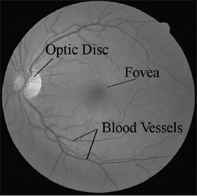
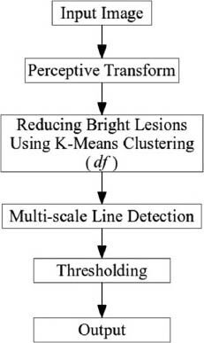
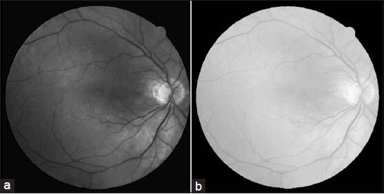
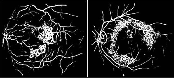
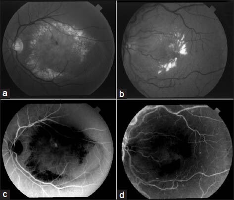
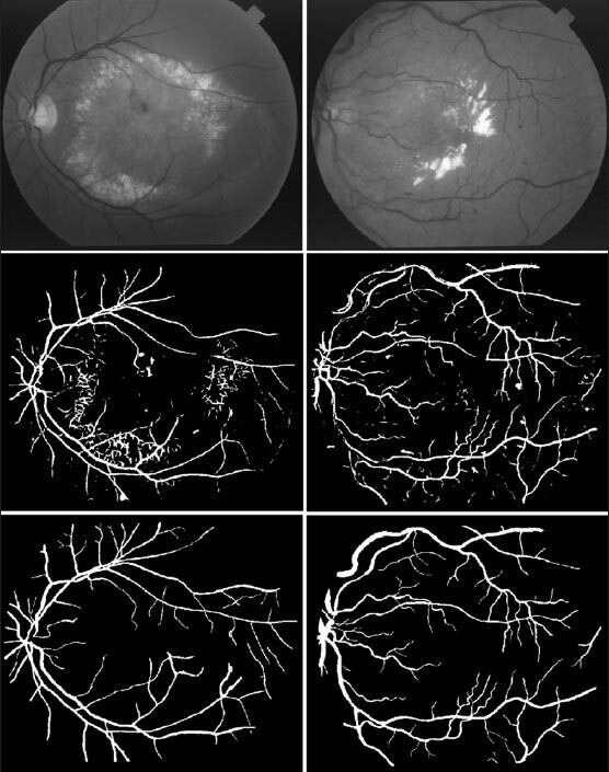
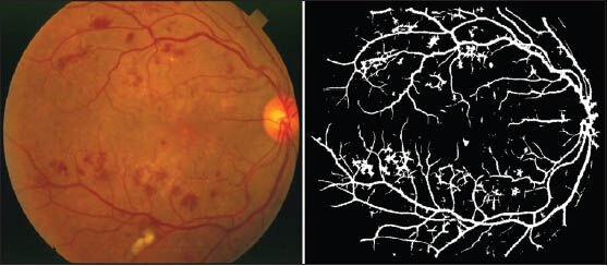
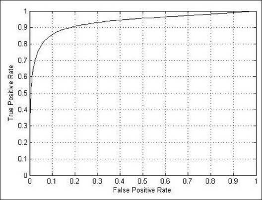
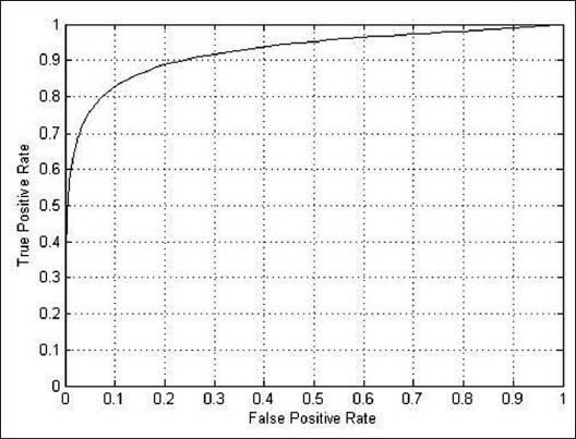
Detecting blood vessels is a vital task in retinal image analysis. The task is more challenging with the presence of bright and dark lesions in retinal images. Here, a method is proposed to detect vessels in both normal and abnormal retinal fundus images based on their linear features. First, the negative impact of bright lesions is reduced by using K-means segmentation in a perceptive space. Then, a multi-scale line operator is utilized to detect vessels while ignoring some of the dark lesions, which have intensity structures different from the line-shaped vessels in the retina. The proposed algorithm is tested on two publicly available STARE and DRIVE databases. The performance of the method is measured by calculating the area under the receiver operating characteristic curve and the segmentation accuracy. The proposed method achieves 0.9483 and 0.9387 localization accuracy against STARE and DRIVE respectively.

摘要

检测血管是视网膜图像分析中的一项重要任务。在视网膜图像中存在明亮和黑暗病变的情况下,这项任务更具挑战性。在此,提出了一种基于线性特征来检测正常和异常眼底视网膜图像中血管的方法。首先,通过在感知空间中使用K均值分割来减少明亮病变的负面影响。然后,利用多尺度线算子来检测血管,同时忽略一些黑暗病变,这些黑暗病变具有与视网膜中线状血管不同的强度结构。所提出的算法在两个公开可用的STARE和DRIVE数据库上进行了测试。该方法的性能通过计算接收器操作特征曲线下的面积和分割精度来衡量。所提出的方法在STARE和DRIVE数据库上分别实现了0.9483和0.9387的定位精度。

https://cdn.ncbi.nlm.nih.gov/pmc/blobs/f06e/3994716/40e4b9c1f7ee/JMSS-4-122-g020.jpg
https://cdn.ncbi.nlm.nih.gov/pmc/blobs/f06e/3994716/be0e7fc47497/JMSS-4-122-g001.jpg
https://cdn.ncbi.nlm.nih.gov/pmc/blobs/f06e/3994716/400b2538bee3/JMSS-4-122-g002.jpg
https://cdn.ncbi.nlm.nih.gov/pmc/blobs/f06e/3994716/83fedbc8b0e5/JMSS-4-122-g003.jpg
https://cdn.ncbi.nlm.nih.gov/pmc/blobs/f06e/3994716/235400032b03/JMSS-4-122-g005.jpg
https://cdn.ncbi.nlm.nih.gov/pmc/blobs/f06e/3994716/061449d9fec3/JMSS-4-122-g006.jpg
https://cdn.ncbi.nlm.nih.gov/pmc/blobs/f06e/3994716/eff5b3a722a0/JMSS-4-122-g010.jpg
https://cdn.ncbi.nlm.nih.gov/pmc/blobs/f06e/3994716/447d3de22279/JMSS-4-122-g011.jpg
https://cdn.ncbi.nlm.nih.gov/pmc/blobs/f06e/3994716/fbec9ba375e5/JMSS-4-122-g012.jpg
https://cdn.ncbi.nlm.nih.gov/pmc/blobs/f06e/3994716/b1d04eac5e90/JMSS-4-122-g013.jpg
https://cdn.ncbi.nlm.nih.gov/pmc/blobs/f06e/3994716/b3e8ae82e78b/JMSS-4-122-g014.jpg
https://cdn.ncbi.nlm.nih.gov/pmc/blobs/f06e/3994716/9da3c005ca97/JMSS-4-122-g018.jpg
https://cdn.ncbi.nlm.nih.gov/pmc/blobs/f06e/3994716/e2713050c2f8/JMSS-4-122-g019.jpg
https://cdn.ncbi.nlm.nih.gov/pmc/blobs/f06e/3994716/40e4b9c1f7ee/JMSS-4-122-g020.jpg

相似文献

1
Vessel Segmentation in Retinal Images Using Multi-scale Line Operator and K-Means Clustering.基于多尺度线算子和K均值聚类的视网膜图像血管分割
J Med Signals Sens. 2014 Apr;4(2):122-9.
2
Scale-space approximated convolutional neural networks for retinal vessel segmentation.用于视网膜血管分割的尺度空间逼近卷积神经网络。
Comput Methods Programs Biomed. 2019 Sep;178:237-246. doi: 10.1016/j.cmpb.2019.06.030. Epub 2019 Jun 29.
3
Unsupervised fuzzy based vessel segmentation in pathological digital fundus images.基于无监督模糊的病理数字眼底图像血管分割。
J Med Syst. 2010 Oct;34(5):849-58. doi: 10.1007/s10916-009-9299-0. Epub 2009 May 9.
4
A new robust method for blood vessel segmentation in retinal fundus images based on weighted line detector and hidden Markov model.基于加权线检测器和隐马尔可夫模型的视网膜眼底图像血管分割新方法。
Comput Methods Programs Biomed. 2020 Apr;187:105231. doi: 10.1016/j.cmpb.2019.105231. Epub 2019 Nov 23.
5
Segmenting retinal vessels with revised top-bottom-hat transformation and flattening of minimum circumscribed ellipse.基于改进的顶底帽变换和平滑最小外接椭圆的视网膜血管分割。
Med Biol Eng Comput. 2019 Jul;57(7):1481-1496. doi: 10.1007/s11517-019-01967-2. Epub 2019 Mar 22.
6
Construction and application of color fundus image segmentation algorithm based on Multi-Scale local combined global enhancement.基于多尺度局部与全局增强相结合的彩色眼底图像分割算法的构建与应用
Pak J Med Sci. 2021;37(6):1595-1599. doi: 10.12669/pjms.37.6-WIT.4848.
7
Iterative Vessel Segmentation of Fundus Images.眼底图像的迭代血管分割
IEEE Trans Biomed Eng. 2015 Jul;62(7):1738-49. doi: 10.1109/TBME.2015.2403295. Epub 2015 Feb 13.
8
Diabetic and Hypertensive Retinopathy Screening in Fundus Images Using Artificially Intelligent Shallow Architectures.利用人工智能浅层架构进行眼底图像中的糖尿病性和高血压性视网膜病变筛查。
J Pers Med. 2021 Dec 23;12(1):7. doi: 10.3390/jpm12010007.
9
Automated techniques for blood vessels segmentation through fundus retinal images: A review.通过眼底视网膜图像进行血管分割的自动化技术:综述
Microsc Res Tech. 2019 Feb;82(2):153-170. doi: 10.1002/jemt.23172. Epub 2019 Jan 5.
10
Blood vessel segmentation in color fundus images based on regional and Hessian features.基于区域和黑塞特征的彩色眼底图像血管分割
Graefes Arch Clin Exp Ophthalmol. 2017 Aug;255(8):1525-1533. doi: 10.1007/s00417-017-3677-y. Epub 2017 May 4.

引用本文的文献

1
EnsembleEdgeFusion: advancing semantic segmentation in microvascular decompression imaging with innovative ensemble techniques.集成边缘融合:利用创新的集成技术推进微血管减压成像中的语义分割
Sci Rep. 2025 May 23;15(1):17892. doi: 10.1038/s41598-025-02470-5.
2
Unsupervised clustering based coronary artery segmentation.基于无监督聚类的冠状动脉分割
BioData Min. 2025 Mar 7;18(1):21. doi: 10.1186/s13040-025-00435-y.
3
MC-UNet: Multimodule Concatenation Based on U-Shape Network for Retinal Blood Vessels Segmentation.MC-UNet:基于 U 形网络的多模块拼接的视网膜血管分割方法。

本文引用的文献

1
Blood vessel segmentation methodologies in retinal images--a survey.视网膜图像中的血管分割方法综述。
Comput Methods Programs Biomed. 2012 Oct;108(1):407-33. doi: 10.1016/j.cmpb.2012.03.009. Epub 2012 Apr 22.
2
General retinal vessel segmentation using regularization-based multiconcavity modeling.基于正则化多凹模型的视网膜血管整体分割。
IEEE Trans Med Imaging. 2010 Jul;29(7):1369-81. doi: 10.1109/TMI.2010.2043259. Epub 2010 Mar 18.
3
Vascular intersection detection in retina fundus images using a new hybrid approach.
Comput Intell Neurosci. 2022 Nov 3;2022:9917691. doi: 10.1155/2022/9917691. eCollection 2022.
4
Blood Vessel Segmentation of Retinal Image Based on Dense-U-Net Network.基于密集U-Net网络的视网膜图像血管分割
Micromachines (Basel). 2021 Nov 29;12(12):1478. doi: 10.3390/mi12121478.
5
A Multiscale Clustering Approach for Non-IID Nominal Data.多尺度聚类方法在非独立同分布标称数据上的应用。
Comput Intell Neurosci. 2021 Oct 11;2021:8993543. doi: 10.1155/2021/8993543. eCollection 2021.
6
Deep Neural Network-Based Semantic Segmentation of Microvascular Decompression Images.基于深度神经网络的微血管减压术图像语义分割
Sensors (Basel). 2021 Feb 7;21(4):1167. doi: 10.3390/s21041167.
7
Efficient BFCN for Automatic Retinal Vessel Segmentation.用于自动视网膜血管分割的高效BFCN
J Ophthalmol. 2020 Sep 17;2020:6439407. doi: 10.1155/2020/6439407. eCollection 2020.
8
Automatic non-proliferative diabetic retinopathy screening system based on color fundus image.基于彩色眼底图像的自动非增生性糖尿病视网膜病变筛查系统。
Biomed Eng Online. 2017 Oct 26;16(1):122. doi: 10.1186/s12938-017-0414-z.
9
Blood Vessel Extraction in Color Retinal Fundus Images with Enhancement Filtering and Unsupervised Classification.基于增强滤波和无监督分类的彩色眼底图像血管提取。
J Healthc Eng. 2017;2017:4897258. doi: 10.1155/2017/4897258. Epub 2017 Aug 3.
10
Structured Learning for 3-D Perivascular Space Segmentation Using Vascular Features.基于血管特征的 3D 血管周围空间分割的结构化学习。
IEEE Trans Biomed Eng. 2017 Dec;64(12):2803-2812. doi: 10.1109/TBME.2016.2638918. Epub 2017 Mar 1.
利用一种新的混合方法进行视网膜眼底图像中的血管交叉检测。
Comput Biol Med. 2010 Jan;40(1):81-9. doi: 10.1016/j.compbiomed.2009.11.004. Epub 2009 Dec 22.
4
A novel vessel segmentation algorithm for pathological retina images based on the divergence of vector fields.一种基于向量场散度的病理性视网膜图像血管分割新算法。
IEEE Trans Med Imaging. 2008 Feb;27(2):237-46. doi: 10.1109/TMI.2007.909827.
5
Detection of blood vessels in retinal images using two-dimensional matched filters.利用二维匹配滤波器检测视网膜图像中的血管。
IEEE Trans Med Imaging. 1989;8(3):263-9. doi: 10.1109/42.34715.
6
Retinal blood vessel segmentation using line operators and support vector classification.使用线算子和支持向量分类的视网膜血管分割
IEEE Trans Med Imaging. 2007 Oct;26(10):1357-65. doi: 10.1109/TMI.2007.898551.
7
Segmentation of blood vessels from red-free and fluorescein retinal images.从无赤光和荧光素视网膜图像中分割血管。
Med Image Anal. 2007 Feb;11(1):47-61. doi: 10.1016/j.media.2006.11.004. Epub 2007 Jan 3.
8
Retinal vessel segmentation using the 2-D Gabor wavelet and supervised classification.使用二维伽柏小波和监督分类进行视网膜血管分割。
IEEE Trans Med Imaging. 2006 Sep;25(9):1214-22. doi: 10.1109/tmi.2006.879967.
9
Segmentation of retinal blood vessels by combining the detection of centerlines and morphological reconstruction.通过结合中心线检测和形态学重建对视网膜血管进行分割。
IEEE Trans Med Imaging. 2006 Sep;25(9):1200-13. doi: 10.1109/tmi.2006.879955.
10
Linear structures in mammographic images: detection and classification.乳腺X线图像中的线性结构:检测与分类
IEEE Trans Med Imaging. 2004 Sep;23(9):1077-86. doi: 10.1109/TMI.2004.828675.