• 文献检索
  • 文档翻译
  • 深度研究
  • 学术资讯
  • Suppr Zotero 插件Zotero 插件
  • 邀请有礼
  • 套餐&价格
  • 历史记录
应用&插件
Suppr Zotero 插件Zotero 插件浏览器插件Mac 客户端Windows 客户端微信小程序
定价
高级版会员购买积分包购买API积分包
服务
文献检索文档翻译深度研究API 文档MCP 服务
关于我们
关于 Suppr公司介绍联系我们用户协议隐私条款
关注我们

Suppr 超能文献

核心技术专利:CN118964589B侵权必究
粤ICP备2023148730 号-1Suppr @ 2026

文献检索

告别复杂PubMed语法,用中文像聊天一样搜索,搜遍4000万医学文献。AI智能推荐,让科研检索更轻松。

立即免费搜索

文件翻译

保留排版,准确专业,支持PDF/Word/PPT等文件格式,支持 12+语言互译。

免费翻译文档

深度研究

AI帮你快速写综述,25分钟生成高质量综述,智能提取关键信息,辅助科研写作。

立即免费体验

蓝色激光成像放大技术与凹陷模式分析对大肠息肉诊断能力的比较

Comparison of the diagnostic ability of blue laser imaging magnification versus pit pattern analysis for colorectal polyps.

作者信息

Nakano Arihiro, Hirooka Yoshiki, Yamamura Takeshi, Watanabe Osamu, Nakamura Masanao, Funasaka Kohei, Ohno Eizaburo, Kawashima Hiroki, Miyahara Ryoji, Goto Hidemi

机构信息

Department of Gastroenterology and Hepatology, Nagoya University Graduate School of Medicine, Showa-ku, Nagoya, Japan.

Department of Endoscopy, Nagoya University Hospital, Showa-ku, Nagoya, Japan.

出版信息

Endosc Int Open. 2017 Apr;5(4):E224-E231. doi: 10.1055/s-0043-102400.

DOI:10.1055/s-0043-102400
PMID:28367494
原文链接:https://pmc.ncbi.nlm.nih.gov/articles/PMC5362373/
Abstract

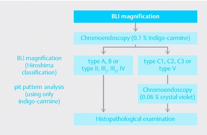
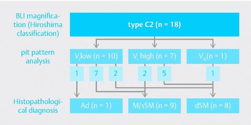
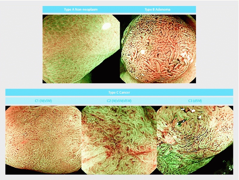
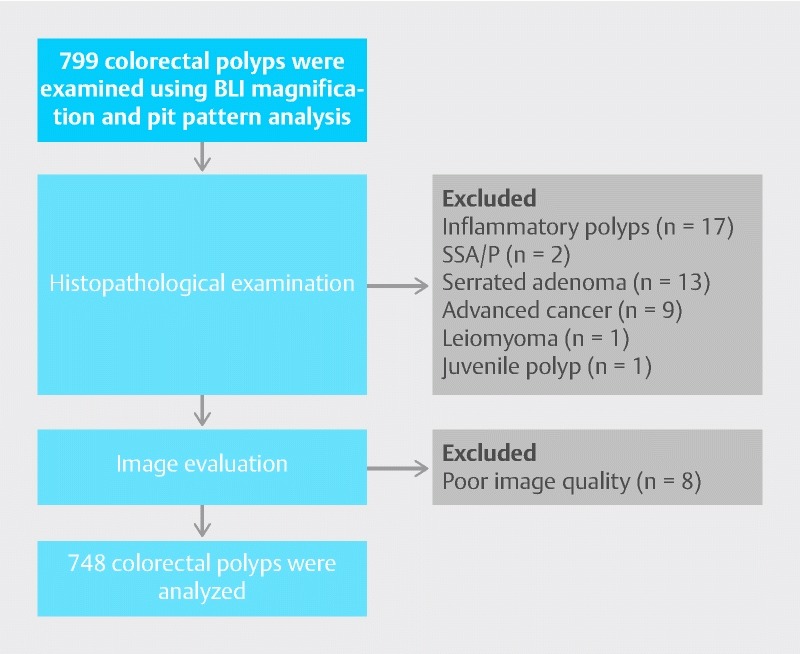
There have been few evaluations of the diagnostic ability of new narrow band light observation blue laser imaging (BLI). The present prospective study compared the diagnostic ability of BLI magnification and pit pattern analysis for colorectal polyps. We collected lesions prospectively, and the analysis of images was made by two endoscopists, retrospectively. A total of 799 colorectal polyps were examined by BLI magnification and pit pattern analysis at Nagoya University Hospital. The Hiroshima narrow-band imaging classification was used for BLI. Differentiation of neoplastic from non-neoplastic lesions and diagnosis of deeply invasive submucosal cancer (dSM) were compared between BLI magnification and pit pattern analysis. Type C2 in the Hiroshima classification was evaluated separately, because application of this category as an index of the depth of cancer invasion was considered difficult. We analyzed 748 colorectal polyps, excluding 51 polyps that were inflammatory polyps, sessile serrated adenoma/polyps, serrated adenomas, advanced colorectal cancers, or other lesions. The accuracy of differential diagnosis between neoplastic and non-neoplastic lesions was 98.4 % using BLI magnification and 98.7 % with pit pattern analysis. In addition, the diagnostic accuracy of BLI magnification and pit pattern analysis for dSM for cancer was 89.5 % and 92.1 %, respectively. When type C2 lesions were excluded, the diagnostic accuracy of BLI for dSM was 95.9 %. The 18 type C2 lesions comprised 1 adenoma, 9 intramucosal or slightly invasive submucosal cancers, and 8 dSM. Pit pattern analysis allowed accurate diagnosis of the depth of invasion in 13 lesions (72.2 %). Most colorectal polyps could be diagnosed accurately by BLI magnification without pit pattern analysis, but we should add pit pattern analysis for type C2 lesions in the Hiroshima classification.

摘要

关于新型窄带光观察蓝色激光成像(BLI)诊断能力的评估较少。本前瞻性研究比较了BLI放大内镜和pit模式分析对大肠息肉的诊断能力。我们前瞻性地收集病变,并由两名内镜医师对图像进行回顾性分析。名古屋大学医院对799例大肠息肉进行了BLI放大内镜和pit模式分析检查。BLI采用广岛窄带成像分类。比较了BLI放大内镜和pit模式分析在鉴别肿瘤性病变与非肿瘤性病变以及诊断深层浸润性黏膜下癌(dSM)方面的差异。广岛分类中的C2型单独进行评估,因为将该类别用作癌症浸润深度的指标被认为存在困难。我们分析了748例大肠息肉,排除了51例炎性息肉、无蒂锯齿状腺瘤/息肉、锯齿状腺瘤、进展期大肠癌或其他病变。使用BLI放大内镜鉴别肿瘤性病变与非肿瘤性病变的诊断准确率为98.4%,pit模式分析为98.7%。此外,BLI放大内镜和pit模式分析对dSM癌的诊断准确率分别为89.5%和92.1%。排除C2型病变后,BLI对dSM的诊断准确率为95.9%。18例C2型病变包括1例腺瘤、9例黏膜内或轻度浸润性黏膜下癌和8例dSM。pit模式分析能够准确诊断13例病变(72.2%)的浸润深度。大多数大肠息肉无需pit模式分析仅通过BLI放大内镜就能准确诊断,但对于广岛分类中的C2型病变,我们应增加pit模式分析。

https://cdn.ncbi.nlm.nih.gov/pmc/blobs/7f2c/5362373/c2149ba64586/10-1055-s-0043-102400-i709ei4.jpg
https://cdn.ncbi.nlm.nih.gov/pmc/blobs/7f2c/5362373/41223b68d8f8/10-1055-s-0043-102400-i709ei1.jpg
https://cdn.ncbi.nlm.nih.gov/pmc/blobs/7f2c/5362373/06d22b5e209c/10-1055-s-0043-102400-i709ei2.jpg
https://cdn.ncbi.nlm.nih.gov/pmc/blobs/7f2c/5362373/376f9e378b0f/10-1055-s-0043-102400-i709ei3.jpg
https://cdn.ncbi.nlm.nih.gov/pmc/blobs/7f2c/5362373/c2149ba64586/10-1055-s-0043-102400-i709ei4.jpg

相似文献

1
Comparison of the diagnostic ability of blue laser imaging magnification versus pit pattern analysis for colorectal polyps.蓝色激光成像放大技术与凹陷模式分析对大肠息肉诊断能力的比较
Endosc Int Open. 2017 Apr;5(4):E224-E231. doi: 10.1055/s-0043-102400.
2
Ability of a novel blue laser imaging system for the diagnosis of colorectal polyps.一种新型蓝色激光成像系统诊断大肠息肉的能力。
Dig Endosc. 2014 Mar;26(2):250-8. doi: 10.1111/den.12127. Epub 2013 Jun 3.
3
The ability of a novel blue laser imaging system for the diagnosis of invasion depth of colorectal neoplasms.新型蓝激光成像系统诊断结直肠肿瘤侵犯深度的能力。
J Gastroenterol. 2014 Jan;49(1):73-80. doi: 10.1007/s00535-013-0772-7. Epub 2013 Mar 15.
4
BASIC (BLI Adenoma Serrated International Classification) classification for colorectal polyp characterization with blue light imaging.基于蓝光成像的结直肠息肉特征的 BASIC(BLI 锯齿状腺瘤国际分类)分类。
Endoscopy. 2018 Mar;50(3):211-220. doi: 10.1055/s-0043-121570. Epub 2017 Oct 24.
5
Evaluation of microvessels in colorectal tumors by narrow band imaging magnification: including comparison with magnifying chromoendoscopy.窄带成像放大内镜评估结直肠肿瘤中的微血管:包括与放大染色内镜的比较。
Dig Dis Sci. 2011 Feb;56(2):532-8. doi: 10.1007/s10620-010-1293-3. Epub 2010 Jun 11.
6
Diagnostic accuracy of narrow-band imaging and pit pattern analysis significantly improved for less-experienced endoscopists after an expanded training program.经过扩展培训计划,经验较少的内镜医生的窄带成像和 pit 模式分析的诊断准确性显著提高。
Gastrointest Endosc. 2010 Jul;72(1):127-35. doi: 10.1016/j.gie.2010.01.054. Epub 2010 May 20.
7
Relationship between narrow-band imaging magnifying observation and pit pattern diagnosis in colorectal tumors.结直肠肿瘤的窄带成像放大观察与pit pattern 诊断的关系。
Digestion. 2013;87(1):53-8. doi: 10.1159/000343940. Epub 2013 Jan 21.
8
Comparison of blue laser imaging and light-emitting diode-blue light imaging for the characterization of colorectal polyps using the Japan narrow-band imaging expert team classification: The LASEREO and ELUXEO COLonoscopic study.使用日本窄带成像专家组分类法比较蓝色激光成像和发光二极管蓝光成像对大肠息肉的特征描述:LASEREO和ELUXEO结肠镜研究。
DEN Open. 2023 May 18;4(1):e245. doi: 10.1002/deo2.245. eCollection 2024 Apr.
9
Usefulness of close observation with non-magnified blue laser imaging for determining cold polypectomy indications.非放大蓝光成像密切观察在确定冷息肉切除术适应证中的应用价值
Scand J Gastroenterol. 2018 Aug;53(8):1013-1017. doi: 10.1080/00365521.2018.1488181. Epub 2018 Jul 24.
10
Pit pattern analysis by magnifying chromoendoscopy for the diagnosis of colorectal polyps.放大色素内镜下的腺管开口形态分析用于结直肠息肉的诊断
J Formos Med Assoc. 2003 Mar;102(3):178-82.

引用本文的文献

1
Improvement of Colonoscopic Image Quality Using a New LED Endoscopic System with Specialized Noise Reduction.使用具有专门降噪功能的新型LED内镜系统改善结肠镜检查图像质量。
Diagnostics (Basel). 2025 Jun 19;15(12):1569. doi: 10.3390/diagnostics15121569.
2
Comparison of LED and LASER Colonoscopy About Linked Color Imaging and Blue Laser/Light Imaging of Colorectal Tumors in a Multinational Study.一项多国研究中 LED 与激光结肠镜检查在大肠肿瘤的联动成像及蓝色激光/光成像方面的比较
Dig Dis Sci. 2023 Oct;68(10):3943-3952. doi: 10.1007/s10620-023-08057-2. Epub 2023 Aug 9.
3
The Efficacy of Tumor Characterization for Colorectal Lesions with Blue Light Imaging of a Compact Light-Emitting Diode Endoscopic System Compared to a Laser Endoscopic System: A Pilot Study.

本文引用的文献

1
Narrow-band imaging (NBI) magnifying endoscopic classification of colorectal tumors proposed by the Japan NBI Expert Team.日本窄带成像(NBI)专家组提出的结直肠肿瘤窄带成像(NBI)放大内镜分类。
Dig Endosc. 2016 Jul;28(5):526-33. doi: 10.1111/den.12644. Epub 2016 Apr 20.
2
Japanese Society for Cancer of the Colon and Rectum (JSCCR) Guidelines 2014 for treatment of colorectal cancer.日本结直肠癌学会(JSCCR)2014年结直肠癌治疗指南。
Int J Clin Oncol. 2015 Apr;20(2):207-39. doi: 10.1007/s10147-015-0801-z. Epub 2015 Mar 18.
3
Real-time optical diagnosis for diminutive colorectal polyps using narrow-band imaging: the VALID randomised clinical trial.
与激光内镜系统相比,紧凑型发光二极管内镜系统蓝光成像对结直肠病变的肿瘤特征识别效能:一项初步研究
Gastroenterol Res Pract. 2022 Apr 12;2022:9998280. doi: 10.1155/2022/9998280. eCollection 2022.
4
Diagnostic performance of endoscopic classifications for neoplastic lesions in patients with ulcerative colitis: A retrospective case-control study.内镜分类对溃疡性结肠炎患者肿瘤性病变的诊断性能:一项回顾性病例对照研究。
World J Gastroenterol. 2022 Mar 14;28(10):1055-1066. doi: 10.3748/wjg.v28.i10.1055.
5
Electronic chromo-endoscopy: technical details and a clinical perspective.电子染色内镜检查:技术细节与临床展望。
Transl Gastroenterol Hepatol. 2022 Jan 25;7:6. doi: 10.21037/tgh-19-373. eCollection 2022.
6
Colorectal Endoscopic Submucosal Dissection: An Update on Best Practice.结直肠内镜黏膜下剥离术:最佳实践的最新进展
Clin Exp Gastroenterol. 2021 Aug 3;14:317-330. doi: 10.2147/CEG.S249869. eCollection 2021.
7
Clinical Applications of Linked Color Imaging and Blue Laser/Light Imaging in the Screening, Diagnosis, and Treatment of Superficial Colorectal Tumors.联合彩色成像和蓝光成像在浅表性结直肠肿瘤筛查、诊断及治疗中的临床应用
Clin Endosc. 2021 Jul;54(4):488-493. doi: 10.5946/ce.2021.157. Epub 2021 Jul 14.
8
Diagnostic efficacy of the Japan Narrow-band-imaging Expert Team and Pit pattern classifications for colorectal lesions: A meta-analysis.日本窄带成像专家组和凹坑模式分类法对结直肠病变的诊断效能:一项荟萃分析。
World J Gastroenterol. 2020 Oct 28;26(40):6279-6294. doi: 10.3748/wjg.v26.i40.6279.
9
The efficacy of tumor characterization and tumor detectability of linked color imaging and blue laser imaging with an LED endoscope compared to a LASER endoscope.与激光内窥镜相比,联用彩色成像和蓝色激光成像的发光二极管内窥镜在肿瘤特征识别及肿瘤可检测性方面的效能。
Int J Colorectal Dis. 2020 May;35(5):815-825. doi: 10.1007/s00384-020-03532-y. Epub 2020 Feb 22.
10
Evaluation of the impact of linked color imaging for improving the visibility of colonic polyp.评估联动彩色成像对提高结肠息肉可视性的影响。
Oncol Lett. 2019 Nov;18(5):5555-5560. doi: 10.3892/ol.2019.10917. Epub 2019 Sep 24.
使用窄带成像的微小结直肠息肉实时光学诊断:VALID 随机临床试验。
Gut. 2015 Oct;64(10):1569-77. doi: 10.1136/gutjnl-2014-307742. Epub 2014 Nov 11.
4
Long-term colorectal-cancer mortality after adenoma removal.腺瘤切除术后的长期结直肠癌死亡率。
N Engl J Med. 2014 Aug 28;371(9):799-807. doi: 10.1056/NEJMoa1315870.
5
Endoscopic prediction of deep submucosal invasive carcinoma: validation of the narrow-band imaging international colorectal endoscopic (NICE) classification.内镜预测黏膜下深层浸润癌:窄带成像国际结直肠内镜(NICE)分类的验证。
Gastrointest Endosc. 2013 Oct;78(4):625-32. doi: 10.1016/j.gie.2013.04.185. Epub 2013 Jul 30.
6
Ability of a novel blue laser imaging system for the diagnosis of colorectal polyps.一种新型蓝色激光成像系统诊断大肠息肉的能力。
Dig Endosc. 2014 Mar;26(2):250-8. doi: 10.1111/den.12127. Epub 2013 Jun 3.
7
The ability of a novel blue laser imaging system for the diagnosis of invasion depth of colorectal neoplasms.新型蓝激光成像系统诊断结直肠肿瘤侵犯深度的能力。
J Gastroenterol. 2014 Jan;49(1):73-80. doi: 10.1007/s00535-013-0772-7. Epub 2013 Mar 15.
8
Relationship between narrow-band imaging magnifying observation and pit pattern diagnosis in colorectal tumors.结直肠肿瘤的窄带成像放大观察与pit pattern 诊断的关系。
Digestion. 2013;87(1):53-8. doi: 10.1159/000343940. Epub 2013 Jan 21.
9
Narrow band imaging to differentiate neoplastic and non-neoplastic colorectal polyps in real time: a meta-analysis of diagnostic operating characteristics.实时窄带成像鉴别结直肠息肉的良恶性:诊断操作特性的荟萃分析。
Gut. 2013 Dec;62(12):1704-13. doi: 10.1136/gutjnl-2012-303965. Epub 2013 Jan 7.
10
Characteristic magnifying narrow-band imaging features of colorectal tumors in each growth type.各生长类型结直肠肿瘤特征性放大窄带成像表现。
Int J Colorectal Dis. 2013 Apr;28(4):459-68. doi: 10.1007/s00384-012-1612-6. Epub 2012 Dec 4.