• 文献检索
  • 文档翻译
  • 深度研究
  • 学术资讯
  • Suppr Zotero 插件Zotero 插件
  • 邀请有礼
  • 套餐&价格
  • 历史记录
应用&插件
Suppr Zotero 插件Zotero 插件浏览器插件Mac 客户端Windows 客户端微信小程序
定价
高级版会员购买积分包购买API积分包
服务
文献检索文档翻译深度研究API 文档MCP 服务
关于我们
关于 Suppr公司介绍联系我们用户协议隐私条款
关注我们

Suppr 超能文献

核心技术专利:CN118964589B侵权必究
粤ICP备2023148730 号-1Suppr @ 2026

文献检索

告别复杂PubMed语法,用中文像聊天一样搜索,搜遍4000万医学文献。AI智能推荐,让科研检索更轻松。

立即免费搜索

文件翻译

保留排版,准确专业,支持PDF/Word/PPT等文件格式,支持 12+语言互译。

免费翻译文档

深度研究

AI帮你快速写综述,25分钟生成高质量综述,智能提取关键信息,辅助科研写作。

立即免费体验

通过光学相干断层扫描血管造影评估的与糖尿病相关的视网膜微血管改变:一项横断面分析。

Retinal microvascular alterations related to diabetes assessed by optical coherence tomography angiography: A cross-sectional analysis.

作者信息

Gozlan Julien, Ingrand Pierre, Lichtwitz Olivier, Cazet-Supervielle Agathe, Benoudis Léa, Boissonnot Michele, Hadjadj Samy, Leveziel Nicolas

机构信息

Department of Ophthalmology, CHU Poitiers Public Health Department, University of Poitiers University of Poitiers, UFR de Médecine et Pharmacie Biostatistics, INSERM CIC 1402, Centre d'investigation clinique, University Hospital of Poitiers Diabetology Department, Poitiers - Coordinating Center of the DIAB2NEPHROGENE Study INSERM 1084, University of Poitiers, France.

出版信息

Medicine (Baltimore). 2017 Apr;96(15):e6427. doi: 10.1097/MD.0000000000006427.

DOI:10.1097/MD.0000000000006427
PMID:28403072
原文链接:https://pmc.ncbi.nlm.nih.gov/articles/PMC5403069/
Abstract

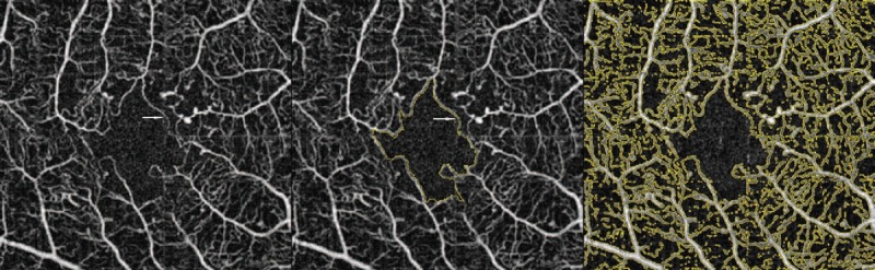
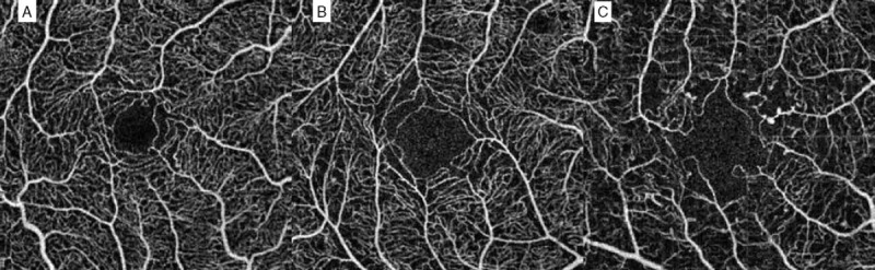
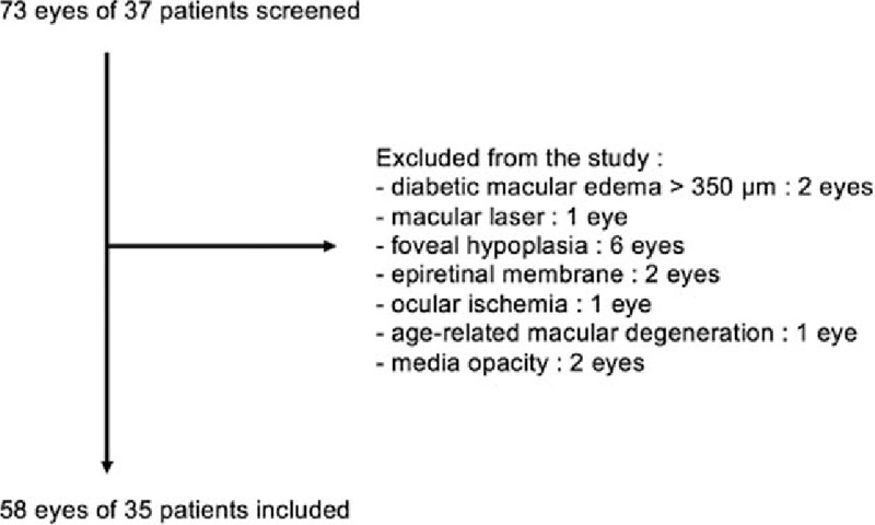
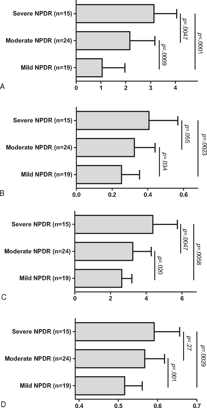
Fluorescein angiography has been so far the gold-standard test to assess diabetic macular ischemia (DMI), a cause of irreversible visual impairment in diabetic patients. The aim of this study was to investigate foveal avascular zone (FAZ) and perifoveal microcirculation changes in eyes with nonproliferative diabetic retinopathy (NPDR) using optical coherence tomography angiography (OCTA), a new and noninvasive vascular imaging technique.Cross-sectional study including eyes of diabetic patients with NPDR.All patients underwent medical history, best-corrected visual acuity (BCVA) measurement, slit-lamp and fundus examination, multicolor imaging, SD-OCT, and swept-source OCT. OCTA was performed in order to assess macular superficial and deep capillary plexus, and swept-source OCT was performed to evaluate the central choroidal thickness.Fifty-eight eyes of 35 patients with a mean age of 61.8 years (±12.1) with mean HbA1C level of 7.6% (±1.5) were included in this study. Among them, 19 eyes had mild NPDR, 24 eyes had moderate NPDR, and 15 eyes had severe NPDR. There was a significant progression between NPDR stages for FAZ grade (P < 0.0001), surface (P = 0.0036) and perimeter (P = 0.0001), and for superficial capillary plexus nonperfusion index (NPI) (P = 0.0009). Moreover, a significant correlation was found between NPI and BCVA (P = 0.007).OCT angiography is a useful noninvasive tool to explore early phases of diabetic retinopathy, which are not routinely explore with fluorescein angiography and not precisely enough with color photographs. NPI and foveal avascular zone parameters are correlated with glycated hemoglobin in patients with NPDR. If confirmed by further studies, these results could represent a mean to sensibilize diabetic patients to their disease.

摘要

荧光素血管造影术一直是评估糖尿病性黄斑缺血(DMI)的金标准检查,DMI是糖尿病患者不可逆视力损害的一个原因。本研究的目的是使用光学相干断层扫描血管造影术(OCTA)这一新型非侵入性血管成像技术,研究非增殖性糖尿病视网膜病变(NPDR)患者眼睛的黄斑无血管区(FAZ)和黄斑周围微循环变化。

横断面研究,纳入患有NPDR的糖尿病患者的眼睛。

所有患者均接受病史采集、最佳矫正视力(BCVA)测量、裂隙灯和眼底检查、多色成像、SD-OCT以及扫频源OCT检查。进行OCTA以评估黄斑浅层和深层毛细血管丛,进行扫频源OCT以评估中心脉络膜厚度。

本研究纳入了35例平均年龄61.8岁(±12.1)、平均糖化血红蛋白(HbA1C)水平为7.6%(±1.5)的患者的58只眼睛。其中,19只眼睛患有轻度NPDR,24只眼睛患有中度NPDR,15只眼睛患有重度NPDR。FAZ分级(P<0.0001)、面积(P=0.0036)和周长(P=0.0001)以及浅层毛细血管丛无灌注指数(NPI)(P=0.0009)在NPDR各阶段之间有显著进展。此外, 发现NPI与BCVA之间存在显著相关性(P=0.007)。

OCT血管造影术是探索糖尿病视网膜病变早期阶段的一种有用的非侵入性工具,而荧光素血管造影术通常不会对这些早期阶段进行检查,彩色照片对其检查又不够精确。NPDR患者的NPI和黄斑无血管区参数与糖化血红蛋白相关。如果进一步的研究证实了这些结果,那么这些结果可能代表了一种提高糖尿病患者对自身疾病认识的手段。

https://cdn.ncbi.nlm.nih.gov/pmc/blobs/c52b/5403069/bce77d57da0b/medi-96-e6427-g007.jpg
https://cdn.ncbi.nlm.nih.gov/pmc/blobs/c52b/5403069/b58567fc03aa/medi-96-e6427-g001.jpg
https://cdn.ncbi.nlm.nih.gov/pmc/blobs/c52b/5403069/7d0b3a30447f/medi-96-e6427-g002.jpg
https://cdn.ncbi.nlm.nih.gov/pmc/blobs/c52b/5403069/253d03be1caa/medi-96-e6427-g006.jpg
https://cdn.ncbi.nlm.nih.gov/pmc/blobs/c52b/5403069/bce77d57da0b/medi-96-e6427-g007.jpg

相似文献

1
Retinal microvascular alterations related to diabetes assessed by optical coherence tomography angiography: A cross-sectional analysis.通过光学相干断层扫描血管造影评估的与糖尿病相关的视网膜微血管改变:一项横断面分析。
Medicine (Baltimore). 2017 Apr;96(15):e6427. doi: 10.1097/MD.0000000000006427.
2
Deep Capillary Macular Perfusion Indices Obtained with OCT Angiography Correlate with Degree of Nonproliferative Diabetic Retinopathy.通过光学相干断层扫描血管造影获得的深部黄斑毛细血管灌注指数与非增殖性糖尿病视网膜病变程度相关。
Eur J Ophthalmol. 2017 Nov 8;27(6):716-729. doi: 10.5301/ejo.5000948.
3
Interaction Between the Distribution of Diabetic Retinopathy Lesions and the Association of Optical Coherence Tomography Angiography Scans With Diabetic Retinopathy Severity.糖尿病视网膜病变病变分布与光学相干断层扫描血管造影扫描与糖尿病视网膜病变严重程度的相关性分析。
JAMA Ophthalmol. 2020 Dec 1;138(12):1291-1297. doi: 10.1001/jamaophthalmol.2020.4516.
4
[Correlation of capillary plexus with visual acuity in idiopathic macular epiretinal membrane eyes using optical coherence tomography angiography].[利用光学相干断层扫描血管造影术研究特发性黄斑视网膜前膜眼中毛细血管丛与视力的相关性]
Zhonghua Yan Ke Za Zhi. 2019 Oct 11;55(10):757-762. doi: 10.3760/cma.j.issn.0412-4081.2019.10.006.
5
Linking Retinal Microvasculature Features With Severity of Diabetic Retinopathy Using Optical Coherence Tomography Angiography.使用光学相干断层扫描血管造影术将视网膜微血管特征与糖尿病性视网膜病变的严重程度相联系。
Invest Ophthalmol Vis Sci. 2016 Jul 1;57(9):OCT519-25. doi: 10.1167/iovs.15-18901.
6
Quantification of Diabetic Macular Ischemia Using Optical Coherence Tomography Angiography and Its Relationship with Visual Acuity.利用光相干断层扫描血管造影术对糖尿病性黄斑水肿进行定量分析及其与视力的关系。
Ophthalmology. 2017 Feb;124(2):235-244. doi: 10.1016/j.ophtha.2016.10.008. Epub 2016 Nov 23.
7
Correlation Analysis between Foveal Avascular Zone and Peripheral Ischemic Index in Diabetic Retinopathy: A Pilot Study.糖尿病视网膜病变中黄斑无血管区与周边缺血指数的相关性分析:一项初步研究
Ophthalmol Retina. 2018 Jan;2(1):46-52. doi: 10.1016/j.oret.2017.05.007. Epub 2017 Jul 26.
8
Evaluation of Automatically Quantified Foveal Avascular Zone Metrics for Diagnosis of Diabetic Retinopathy Using Optical Coherence Tomography Angiography.基于光学相干断层扫描血管造影的自动量化中心凹无血管区指标对糖尿病视网膜病变的诊断评估。
Invest Ophthalmol Vis Sci. 2018 May 1;59(6):2212-2221. doi: 10.1167/iovs.17-23498.
9
Optical coherence tomography angiography analysis of foveal microvascular changes and inner retinal layer thinning in patients with diabetes.糖尿病患者黄斑区微血管改变及内层视网膜变薄的光相干断层扫描血管造影分析。
Br J Ophthalmol. 2018 Sep;102(9):1226-1231. doi: 10.1136/bjophthalmol-2017-311149. Epub 2017 Dec 19.
10
Intereye Microvascular Differences in Patients With Same-Stage Diabetic Retinopathy Revealed by OCTA.OCTA 揭示同阶段糖尿病视网膜病变患者的眼内微血管差异。
Invest Ophthalmol Vis Sci. 2024 Jun 3;65(6):11. doi: 10.1167/iovs.65.6.11.

引用本文的文献

1
Comparison of the Usefulness of Optical Coherence Tomography Angiography and Fluorescein Angiography in the Diagnosis of Diabetic Macular Edema.光学相干断层扫描血管造影与荧光素血管造影在糖尿病性黄斑水肿诊断中的应用价值比较
Diagnostics (Basel). 2025 Jul 25;15(15):1873. doi: 10.3390/diagnostics15151873.
2
Ultrastructural imaging biomarkers in diabetic macular edema: A major review.糖尿病性黄斑水肿中的超微结构成像生物标志物:一项主要综述。
Indian J Ophthalmol. 2025 Jan 1;73(Suppl 1):S7-S23. doi: 10.4103/IJO.IJO_878_24. Epub 2024 Dec 24.
3
Relationship between nonperfusion area from widefield optical coherence tomography angiography and macular vascular parameters in diabetic retinopathy.

本文引用的文献

1
Intravitreal Aflibercept for Diabetic Macular Edema: 100-Week Results From the VISTA and VIVID Studies.玻璃体内注射阿柏西普治疗糖尿病性黄斑水肿:VISTA 和 VIVID 研究的 100 周结果。
Ophthalmology. 2015 Oct;122(10):2044-52. doi: 10.1016/j.ophtha.2015.06.017. Epub 2015 Jul 18.
2
IN VIVO CHARACTERIZATION OF RETINAL VASCULARIZATION MORPHOLOGY USING OPTICAL COHERENCE TOMOGRAPHY ANGIOGRAPHY.使用光学相干断层扫描血管造影术对视网膜血管化形态进行体内表征
Retina. 2015 Nov;35(11):2196-203. doi: 10.1097/IAE.0000000000000635.
3
Optical Coherence Tomography Angiography in Diabetic Retinopathy: A Prospective Pilot Study.
广角光相干断层扫描血管造影的无灌注区与糖尿病视网膜病变的黄斑血管参数的关系。
Int Ophthalmol. 2023 Dec;43(12):4803-4814. doi: 10.1007/s10792-023-02882-0. Epub 2023 Oct 18.
4
Assessing the Sensitivity of OCT-A Retinal Vasculature Metrics.评估 OCT-A 视网膜血管测量指标的敏感性。
Transl Vis Sci Technol. 2023 Aug 1;12(8):2. doi: 10.1167/tvst.12.8.2.
5
Effects of smoking on the retina of patients with dry age-related macular degeneration by optical coherence tomography angiography.光学相干断层扫描血管造影观察吸烟对干性年龄相关性黄斑变性患者视网膜的影响。
BMC Ophthalmol. 2022 Jul 22;22(1):315. doi: 10.1186/s12886-022-02525-5.
6
Contribution of optical coherence tomography angiography OCT-A in diabetic maculopathy.光学相干断层扫描血管造影术(OCT-A)在糖尿病性黄斑病变中的作用。
Ann Med Surg (Lond). 2021 Oct 1;70:102904. doi: 10.1016/j.amsu.2021.102904. eCollection 2021 Oct.
7
Associations between Macular OCT Angiography and Nonproliferative Diabetic Retinopathy in Young Patients with Type 1 Diabetes Mellitus.黄斑 OCT 血管造影与年轻 1 型糖尿病患者非增殖性糖尿病视网膜病变的相关性。
J Diabetes Res. 2020 Nov 30;2020:8849116. doi: 10.1155/2020/8849116. eCollection 2020.
8
Optical Coherence Tomography Angiography in Type 1 Diabetes Mellitus. Report 1: Diabetic Retinopathy.1型糖尿病的光学相干断层扫描血管造影。报告1:糖尿病视网膜病变。
Transl Vis Sci Technol. 2020 Sep 30;9(10):34. doi: 10.1167/tvst.9.10.34. eCollection 2020 Sep.
9
Correlations Between Optical Coherence Tomography Angiography Parameters and the Visual Acuity in Patients with Diabetic Retinopathy.糖尿病视网膜病变患者光学相干断层扫描血管造影参数与视力的相关性
Clin Ophthalmol. 2020 Apr 23;14:1107-1115. doi: 10.2147/OPTH.S248881. eCollection 2020.
10
Optical coherence tomography angiography findings of type 1 diabetic patients with diabetic retinopathy, in comparison with type 2 patients.1 型糖尿病患者伴糖尿病视网膜病变与 2 型糖尿病患者的光学相干断层扫描血管造影表现比较。
Graefes Arch Clin Exp Ophthalmol. 2020 Feb;258(2):281-288. doi: 10.1007/s00417-019-04517-6. Epub 2019 Dec 12.
光学相干断层扫描血管造影在糖尿病视网膜病变中的应用:一项前瞻性试点研究。
Am J Ophthalmol. 2015 Jul;160(1):35-44.e1. doi: 10.1016/j.ajo.2015.04.021. Epub 2015 Apr 18.
4
Evaluation of ischemic diabetic maculopathy with Fourier-domain optical coherence tomography and microperimetry.利用傅里叶域光学相干断层扫描和微视野计评估缺血性糖尿病性黄斑病变
Can J Ophthalmol. 2015 Feb;50(1):44-8. doi: 10.1016/j.jcjo.2014.08.005.
5
Diabetic macular ischaemia is associated with narrower retinal arterioles in patients with type 2 diabetes.糖尿病性黄斑缺血与2型糖尿病患者较窄的视网膜小动脉有关。
Acta Ophthalmol. 2015 Feb;93(1):e45-51. doi: 10.1111/aos.12519.
6
Retinal vascular layers in macular telangiectasia type 2 imaged by optical coherence tomographic angiography.光学相干断层扫描血管造影成像观察 2 型黄斑毛细血管扩张症的视网膜血管层。
JAMA Ophthalmol. 2015 Jan;133(1):66-73. doi: 10.1001/jamaophthalmol.2014.3950.
7
Retinal vascular layers imaged by fluorescein angiography and optical coherence tomography angiography.荧光素血管造影和光相干断层扫描血管造影成像的视网膜血管层。
JAMA Ophthalmol. 2015 Jan;133(1):45-50. doi: 10.1001/jamaophthalmol.2014.3616.
8
Optical coherence tomography angiography of optic disc perfusion in glaucoma.青光眼视盘血流的光学相干断层扫描血管造影。
Ophthalmology. 2014 Jul;121(7):1322-32. doi: 10.1016/j.ophtha.2014.01.021. Epub 2014 Mar 12.
9
The effects of macular ischemia on visual acuity in diabetic retinopathy.糖尿病视网膜病变中黄斑缺血对视敏度的影响。
Invest Ophthalmol Vis Sci. 2013 Mar 28;54(3):2353-60. doi: 10.1167/iovs.12-11103.
10
Analysis of foveal avascular zone for grading of diabetic retinopathy severity based on curvelet transform.基于曲波变换的黄斑无血管区分析在糖尿病视网膜病变严重程度分级中的应用。
Graefes Arch Clin Exp Ophthalmol. 2012 Nov;250(11):1607-14. doi: 10.1007/s00417-012-2093-6. Epub 2012 Jul 4.