Ariji Yoshiko, Ariji Eiichiro
Department of Oral and Maxillofacial Radiology, Aichi-Gakuin University School of Dentistry, Nagoya, Japan.
Jpn Dent Sci Rev. 2017 Feb;53(1):11-17. doi: 10.1016/j.jdsr.2016.05.001. Epub 2016 Sep 6.
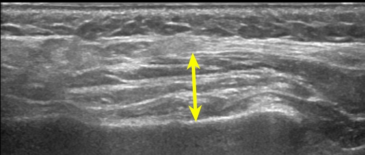
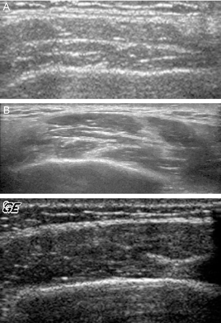
This article reviews recently published studies investigating the MRI and sonographic diagnosis of masticatory muscle myalgia in temporomandibular disorder patients. The MRI and sonographic features of muscle after treatment are also discussed. Literature published within the last 15 years was obtained from the PubMed database using the following Mesh terms: magnetic resonance imaging (MRI) or sonography, masticatory muscle pain, and treatment. MRI and sonography enable accurate visualization and evaluation of the masticatory muscles, thereby increasing our understanding of pathology and cause of pain associated with these muscles. Although therapeutic efficacy is often evaluated based on clinical findings, MR and sonographic imaging studies may also be valuable.
本文综述了近期发表的关于颞下颌关节紊乱症患者咀嚼肌肌痛的MRI和超声诊断的研究。还讨论了治疗后肌肉的MRI和超声特征。使用以下医学主题词从PubMed数据库中获取了过去15年内发表的文献:磁共振成像(MRI)或超声检查、咀嚼肌疼痛和治疗。MRI和超声检查能够准确显示和评估咀嚼肌,从而加深我们对这些肌肉相关病理和疼痛原因的理解。尽管治疗效果通常基于临床发现进行评估,但MR和超声成像研究也可能具有价值。