National Institute for Public Health and the Environment (RIVM) , 3721 MA Bilthoven, The Netherlands.
Department of Environment and Health, VU University , 1081 HV Amsterdam, The Netherlands.
ACS Nano. 2017 May 23;11(5):4542-4552. doi: 10.1021/acsnano.6b08551. Epub 2017 Apr 26.
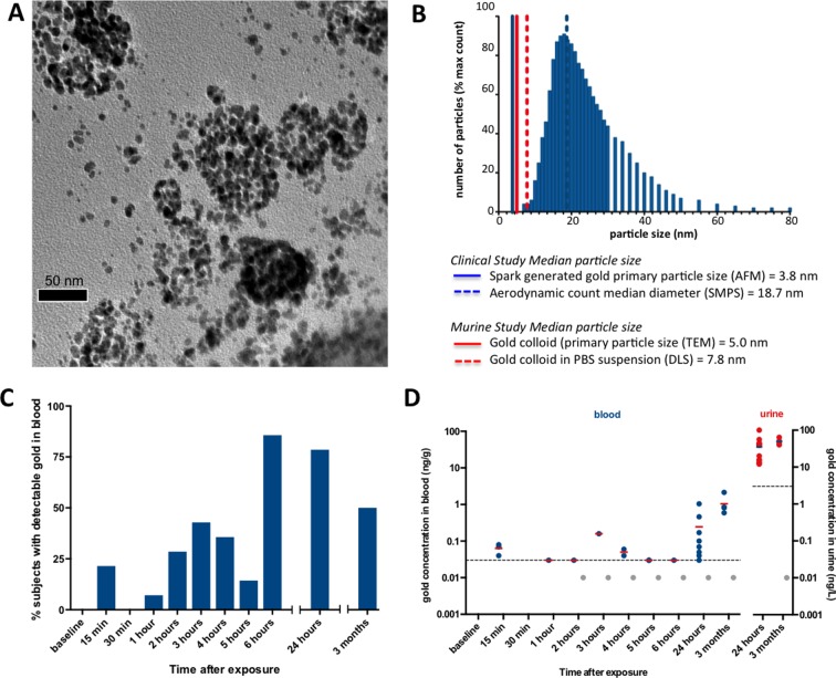
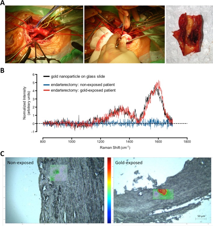
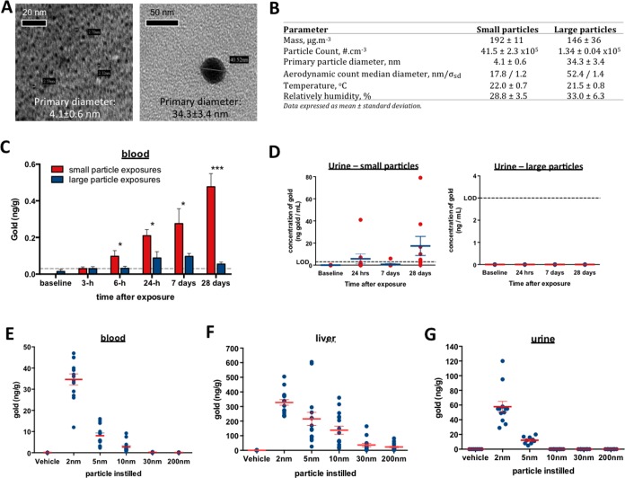
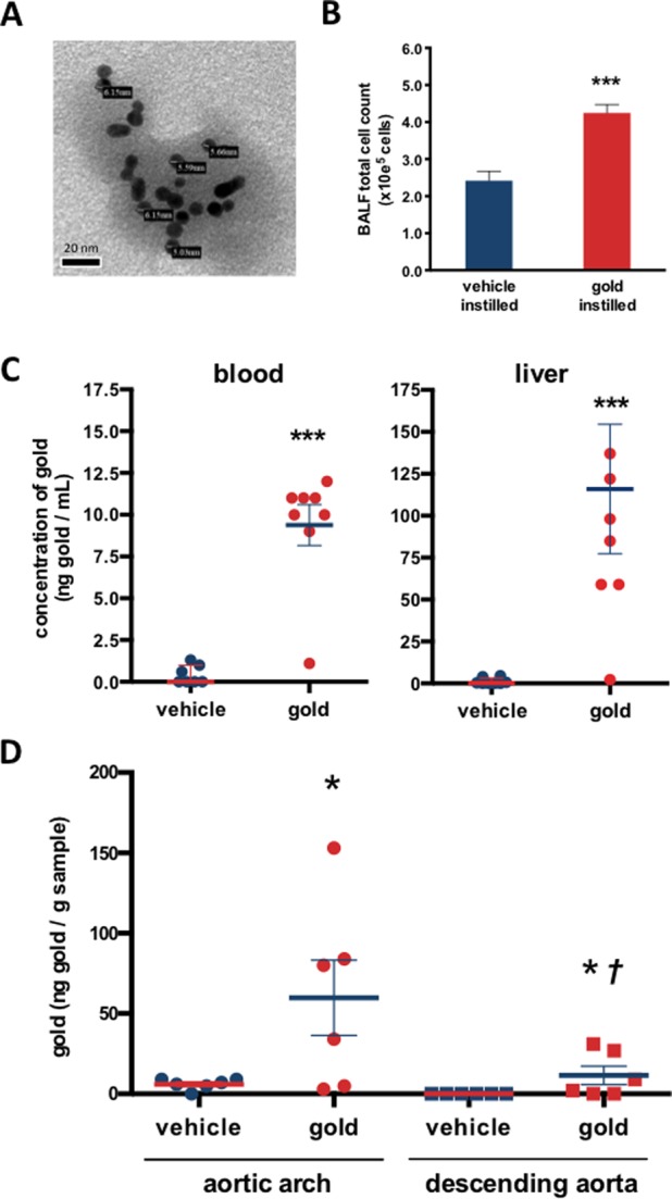
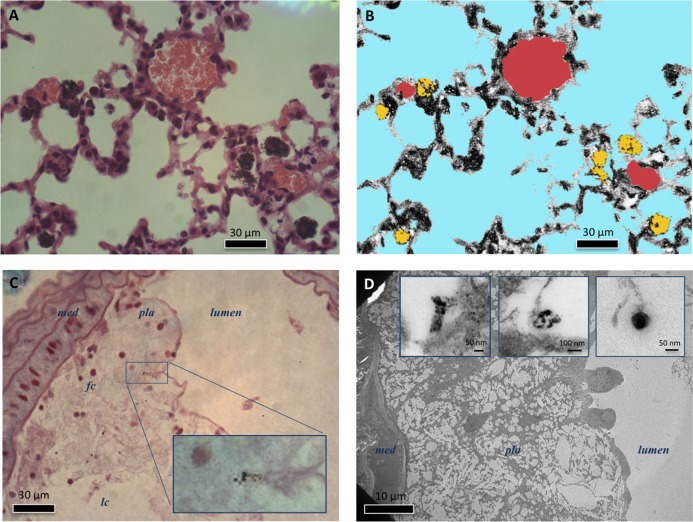
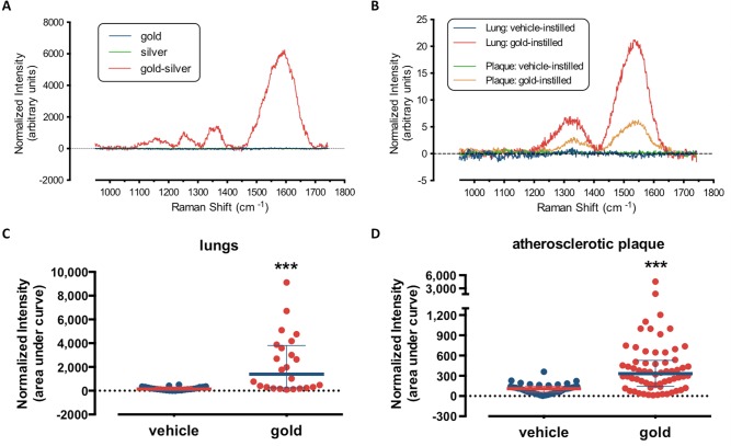
The development of engineered nanomaterials is growing exponentially, despite concerns over their potential similarities to environmental nanoparticles that are associated with significant cardiorespiratory morbidity and mortality. The mechanisms through which inhalation of nanoparticles could trigger acute cardiovascular events are emerging, but a fundamental unanswered question remains: Do inhaled nanoparticles translocate from the lung in man and directly contribute to the pathogenesis of cardiovascular disease? In complementary clinical and experimental studies, we used gold nanoparticles to evaluate particle translocation, permitting detection by high-resolution inductively coupled mass spectrometry and Raman microscopy. Healthy volunteers were exposed to nanoparticles by acute inhalation, followed by repeated sampling of blood and urine. Gold was detected in the blood and urine within 15 min to 24 h after exposure, and was still present 3 months after exposure. Levels were greater following inhalation of 5 nm (primary diameter) particles compared to 30 nm particles. Studies in mice demonstrated the accumulation in the blood and liver following pulmonary exposure to a broader size range of gold nanoparticles (2-200 nm primary diameter), with translocation markedly greater for particles <10 nm diameter. Gold nanoparticles preferentially accumulated in inflammation-rich vascular lesions of fat-fed apolipoproteinE-deficient mice. Furthermore, following inhalation, gold particles could be detected in surgical specimens of carotid artery disease from patients at risk of stroke. Translocation of inhaled nanoparticles into the systemic circulation and accumulation at sites of vascular inflammation provides a direct mechanism that can explain the link between environmental nanoparticles and cardiovascular disease and has major implications for risk management in the use of engineered nanomaterials.
尽管人们对工程纳米材料与环境纳米颗粒的潜在相似性表示担忧,这些颗粒与严重的心肺发病率和死亡率有关,但工程纳米材料的发展仍呈指数级增长。吸入纳米颗粒引发急性心血管事件的机制正在出现,但一个基本的未解决的问题仍然存在:吸入的纳米颗粒是否会从肺部转移到人体中,并直接导致心血管疾病的发病机制?在互补的临床和实验研究中,我们使用金纳米颗粒来评估颗粒转移,允许通过高分辨率电感耦合质谱和拉曼显微镜进行检测。健康志愿者通过急性吸入暴露于纳米颗粒,随后反复采集血液和尿液样本。暴露后 15 分钟至 24 小时内即可在血液和尿液中检测到金,暴露后 3 个月仍可检测到。与 30nm 颗粒相比,吸入 5nm(初级直径)颗粒后血液和尿液中的金含量更高。在小鼠研究中,在肺部暴露于更广泛粒径范围的金纳米颗粒(2-200nm 初级直径)后,血液和肝脏中的积累表明,直径<10nm 的颗粒转移明显更大。金纳米颗粒优先在富含炎症的血管病变中积累,这些病变存在于高脂肪喂养的载脂蛋白 E 缺陷小鼠中。此外,在吸入后,可以在有中风风险的患者的颈动脉疾病手术标本中检测到金颗粒。吸入的纳米颗粒向全身循环的转移和在血管炎症部位的积累提供了一个直接的机制,可以解释环境纳米颗粒与心血管疾病之间的联系,并对工程纳米材料的风险管理具有重要意义。