Conforto Adriana Bastos, Chaim Khallil Taverna, Peres Mario Fernando Prieto, Gonçalves André Leite, Siqueira Inara Laurindo, Barreiros Maria Angela Maramaldo, Amaro Edson
Hospital Israelita Albert Einstein, São Paulo, SP, Brazil.
Einstein (Sao Paulo). 2017 Jan-Mar;15(1):17-23. doi: 10.1590/S1679-45082017AO3719.
To assess changes in blood-oxygen-level-dependent activity after light deprivation compared to regular light exposure in subjects with migraine in the interictal state and in controls.
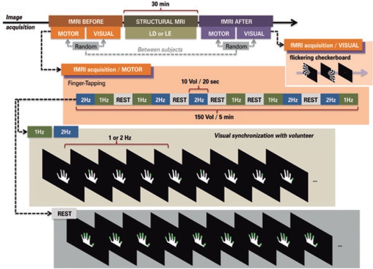
Ten subjects with migraine and ten controls participated in two sessions of functional magnetic resonance imaging. In each session, they performed a finger-tapping task with the right hand, cued by visual stimuli. They were scanned before and after 30 minutes of light deprivation or light exposure. In subjects with migraine, functional magnetic resonance imaging was performed interictally. Analysis of variance was made with the factors time (before or after), session (light deprivation or exposure), and group (migraine or control).
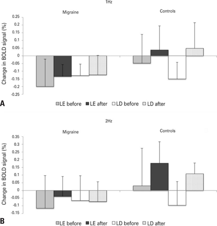
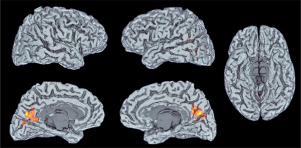
There were significant "group" effects in a cluster in the bilateral cuneus encompassing the superior border of the calcarine sulcus and extrastriate cortex. There were no significant effects of "time", "session", or interactions between these factors.
The main result of this study is consistent with aberrant interictal processing of visual information in migraine. Light deprivation did not modulate functional magnetic resonance imaging activity in subjects with or without migraine.
Avaliar mudanças na atividade cerebral por meio de ressonância magnética funcional após privação luminosa comparada à exposição à luz, em indivíduos com enxaqueca no estado interictal e em controles.
MÉTODOS: Dez indivíduos com enxaqueca e dez controles participaram de duas sessões de ressonância magnética funcional. Em cada sessão, realizaram uma tarefa motora com a mão direita guiada por estímulos visuais. Foram colhidas imagens antes e após 30 minutos de privação luminosa ou exposição à luz. Em indivíduos com enxaqueca, a ressonância funcional foi realizada no período interictal. Foi feita a análise de variância com fatores tempo (antes ou depois), sessão (privação ou exposição à luz) e grupo (enxaqueca ou controle).
Houve efeitos significativos de "grupo" em uma área no cúneo bilateral, incluindo a borda superior do sulco calcarino e o córtex extraestriado. Não houve efeitos significativos de "tempo", "sessão" ou interações entre estes fatores.
CONCLUSÃO: O principal resultado deste estudo sugere um processamento interictal anormal das informações visuais em indivíduos com enxaqueca. A privação luminosa não modulou a atividade na ressonância magnética funcional em indivíduos com ou sem enxaqueca.
评估发作间期偏头痛患者及对照组在经历光剥夺后与正常光照相比,血氧水平依赖性功能磁共振成像活动的变化。
10名偏头痛患者和10名对照者参与了两期功能磁共振成像检查。在每期检查中,他们在视觉刺激提示下用右手执行一项手指敲击任务。在30分钟的光剥夺或光照前后对他们进行扫描。对于偏头痛患者,在发作间期进行功能磁共振成像检查。对时间(之前或之后)、检查期(光剥夺或光照)和组别(偏头痛或对照)等因素进行方差分析。
在双侧楔叶中一个包含距状沟上缘和纹外皮质的区域出现了显著的“组别”效应。“时间”“检查期”或这些因素之间的交互作用均无显著效应。
本研究的主要结果与偏头痛发作间期视觉信息处理异常一致。光剥夺并未调节有或无偏头痛患者的功能磁共振成像活动。
评估发作间期偏头痛患者及对照组在经历光剥夺后与正常光照相比,通过功能磁共振成像测量的大脑活动变化。
10名偏头痛患者和10名对照者参与了两期功能磁共振成像检查。在每期检查中,他们在视觉刺激提示下用右手执行一项运动任务。在30分钟的光剥夺或光照前后采集图像。对于偏头痛患者,在发作间期进行功能磁共振成像检查。对时间(之前或之后)、检查期(光剥夺或光照)和组别(偏头痛或对照)等因素进行方差分析。
在双侧楔叶中一个区域出现了显著的“组别”效应,该区域包括距状沟上缘和纹外皮质。“时间”“检查期”或这些因素之间的交互作用均无显著效应。
本研究的主要结果表明偏头痛患者发作间期视觉信息处理异常。光剥夺并未调节有或无偏头痛患者的功能磁共振成像活动。