Reverchon Flora, Mortaud Stéphane, Sivoyon Maëliss, Maillet Isabelle, Laugeray Anthony, Palomo Jennifer, Montécot Céline, Herzine Améziane, Meme Sandra, Meme William, Erard François, Ryffel Bernhard, Menuet Arnaud, Quesniaux Valérie F J
CNRS, UMR7355, Orleans, France.
Experimental and Molecular Immunology and Neurogenetics, University of Orleans, Orleans, France.
PLoS Pathog. 2017 Apr 27;13(4):e1006322. doi: 10.1371/journal.ppat.1006322. eCollection 2017 Apr.
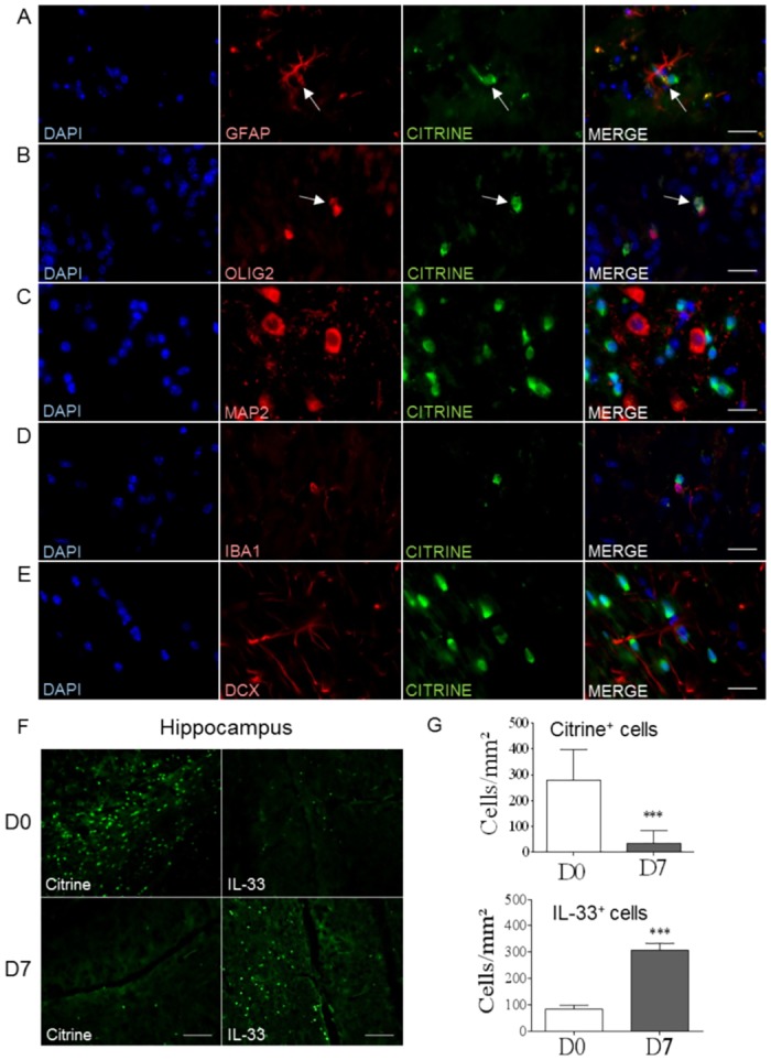
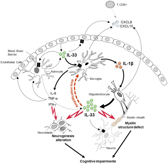
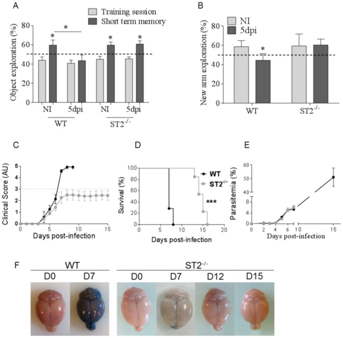
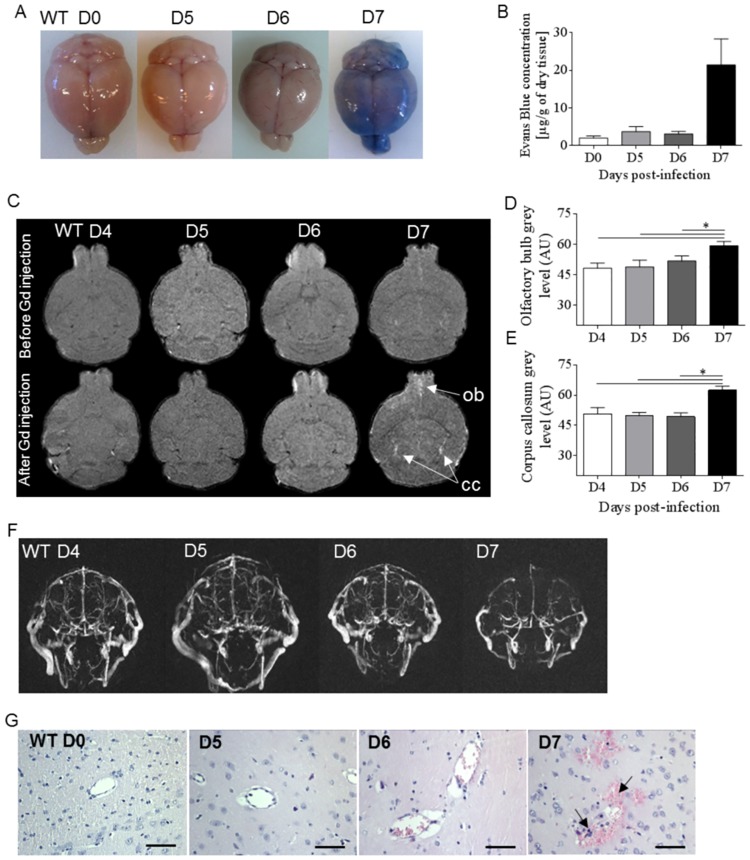
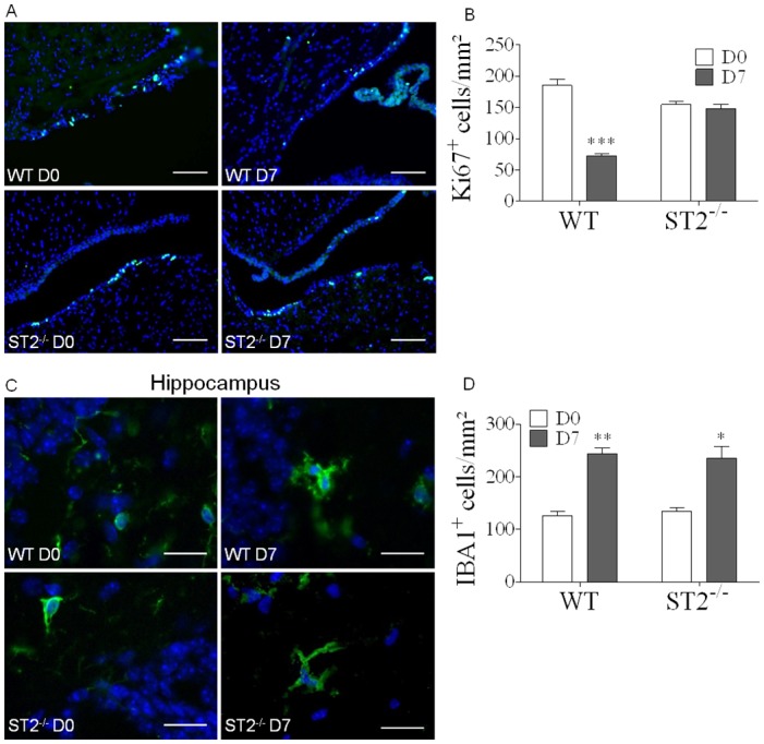
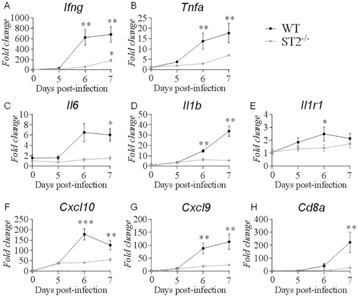
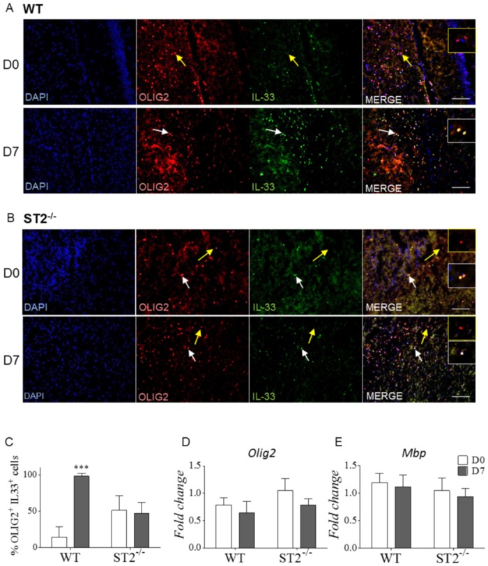
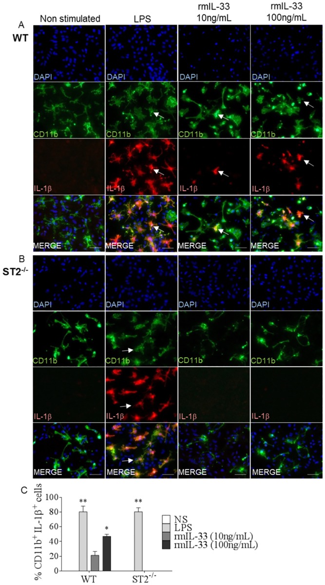
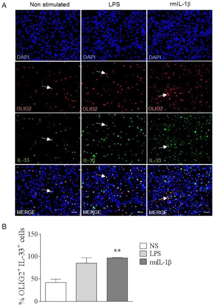
Cerebral malaria (CM) is associated with a high mortality rate and long-term neurocognitive impairment in survivors. The murine model of experimental cerebral malaria (ECM) induced by Plasmodium berghei ANKA (PbA)-infection reproduces several of these features. We reported recently increased levels of IL-33 protein in brain undergoing ECM and the involvement of IL-33/ST2 pathway in ECM development. Here we show that PbA-infection induced early short term and spatial memory defects, prior to blood brain barrier (BBB) disruption, in wild-type mice, while ST2-deficient mice did not develop cognitive defects. PbA-induced neuroinflammation was reduced in ST2-deficient mice with low Ifng, Tnfa, Il1b, Il6, CXCL9, CXCL10 and Cd8a expression, associated with an absence of neurogenesis defects in hippocampus. PbA-infection triggered a dramatic increase of IL-33 expression by oligodendrocytes, through ST2 pathway. In vitro, IL-33/ST2 pathway induced microglia expression of IL-1β which in turn stimulated IL-33 expression by oligodendrocytes. These results highlight the IL-33/ST2 pathway ability to orchestrate microglia and oligodendrocytes responses at an early stage of PbA-infection, with an amplification loop between IL-1β and IL-33, responsible for an exacerbated neuroinflammation context and associated neurological and cognitive defects.
脑型疟疾(CM)与高死亡率以及幸存者的长期神经认知障碍相关。由伯氏疟原虫ANKA(PbA)感染诱导的实验性脑型疟疾(ECM)小鼠模型再现了其中的一些特征。我们最近报道了在发生ECM的大脑中IL-33蛋白水平升高以及IL-33/ST2通路参与ECM的发展。在此我们表明,在野生型小鼠中,PbA感染在血脑屏障(BBB)破坏之前就诱导了早期短期和空间记忆缺陷,而ST2缺陷小鼠未出现认知缺陷。在ST2缺陷小鼠中,PbA诱导的神经炎症减轻,Ifng、Tnfa、Il1b、Il6、CXCL9、CXCL10和Cd8a表达降低,这与海马体中不存在神经发生缺陷有关。PbA感染通过ST2通路触发少突胶质细胞IL-33表达急剧增加。在体外,IL-33/ST2通路诱导小胶质细胞表达IL-1β,进而刺激少突胶质细胞表达IL-33。这些结果突出了IL-33/ST2通路在PbA感染早期协调小胶质细胞和少突胶质细胞反应的能力,以及IL-1β和IL-33之间的放大环路,该环路导致神经炎症加剧以及相关的神经和认知缺陷。