Milbredt Sarah, Waldminghaus Torsten
Chromosome Biology Group, LOEWE Center for Synthetic Microbiology, SYNMIKRO, Philipps-Universität Marburg, D-35043 Marburg, Germany.
Chromosome Biology Group, LOEWE Center for Synthetic Microbiology, SYNMIKRO, Philipps-Universität Marburg, D-35043 Marburg, Germany
G3 (Bethesda). 2017 Jun 7;7(6):1969-1977. doi: 10.1534/g3.117.040782.
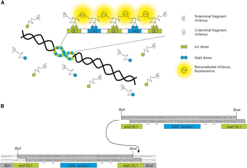
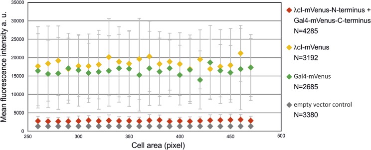
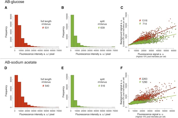
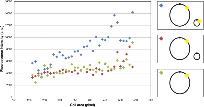
Fluorescence-based methods are widely used to analyze elementary cell processes such as DNA replication or chromosomal folding and segregation. Labeling DNA with a fluorescent protein allows the visualization of its temporal and spatial organization. One popular approach is FROS (fluorescence repressor operator system). This method specifically labels DNA through binding of a fusion of a fluorescent protein and a repressor protein to an operator array, which contains numerous copies of the repressor binding site integrated into the genomic site of interest. Bound fluorescent proteins are then visible as foci in microscopic analyses and can be distinguished from the background fluorescence caused by unbound fusion proteins. Even though this method is widely used, no attempt has been made so far to decrease the background fluorescence to facilitate analysis of the actual signal of interest. Here, we present a new method that greatly reduces the background signal of FROS. BiFCROS (Bimolecular Fluorescence Complementation and Repressor Operator System) is based on fusions of repressor proteins to halves of a split fluorescent protein. Binding to a hybrid FROS array results in fluorescence signals due to bimolecular fluorescence complementation. Only proteins bound to the hybrid FROS array fluoresce, greatly improving the signal to noise ratio compared to conventional FROS. We present the development of BiFCROS and discuss its potential to be used as a fast and single-cell readout for copy numbers of genetic loci.
基于荧光的方法被广泛用于分析基本的细胞过程,如DNA复制、染色体折叠和分离。用荧光蛋白标记DNA可以可视化其时间和空间组织。一种流行的方法是FROS(荧光阻遏物操纵子系统)。该方法通过将荧光蛋白和阻遏蛋白的融合体与操纵子阵列结合来特异性标记DNA,该操纵子阵列包含多个整合到感兴趣基因组位点的阻遏蛋白结合位点拷贝。然后,在显微镜分析中,结合的荧光蛋白可见为焦点,并且可以与未结合的融合蛋白引起的背景荧光区分开来。尽管该方法被广泛使用,但迄今为止尚未尝试降低背景荧光以促进对实际感兴趣信号的分析。在这里,我们提出了一种新方法,该方法大大降低了FROS的背景信号。BiFCROS(双分子荧光互补和阻遏物操纵子系统)基于阻遏蛋白与分裂荧光蛋白的两半的融合。由于双分子荧光互补,与杂交FROS阵列的结合导致荧光信号。只有与杂交FROS阵列结合的蛋白质会发出荧光,与传统FROS相比,大大提高了信噪比。我们介绍了BiFCROS的开发,并讨论了其作为遗传位点拷贝数的快速单细胞读数的潜力。