Mykhaylyk V B, Wagner A, Kraus H
Diamond Light Source, Harwell Campus, Didcot OX11 0DE, UK.
Department of Physics, University of Oxford, Denys Wilkinson Building, Keble Road, Oxford OX1 3RH, UK.
J Synchrotron Radiat. 2017 May 1;24(Pt 3):636-645. doi: 10.1107/S1600577517003484. Epub 2017 Apr 4.
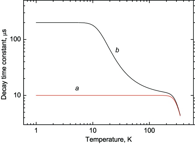
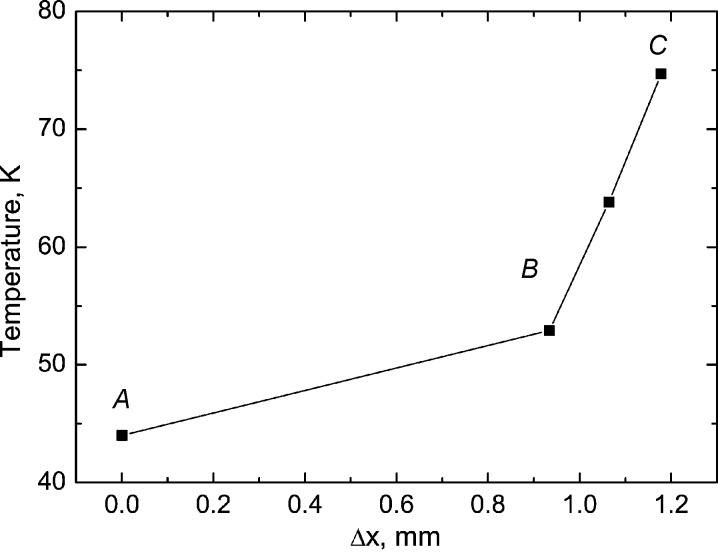
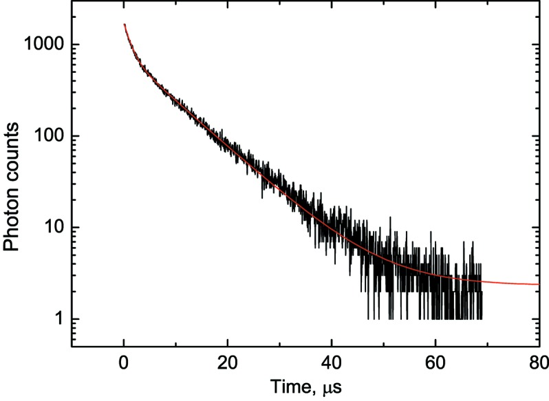
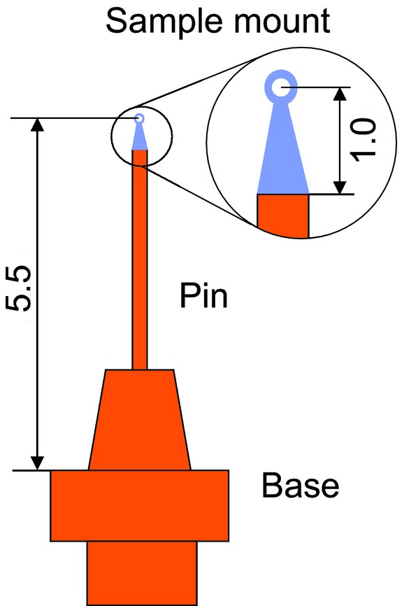
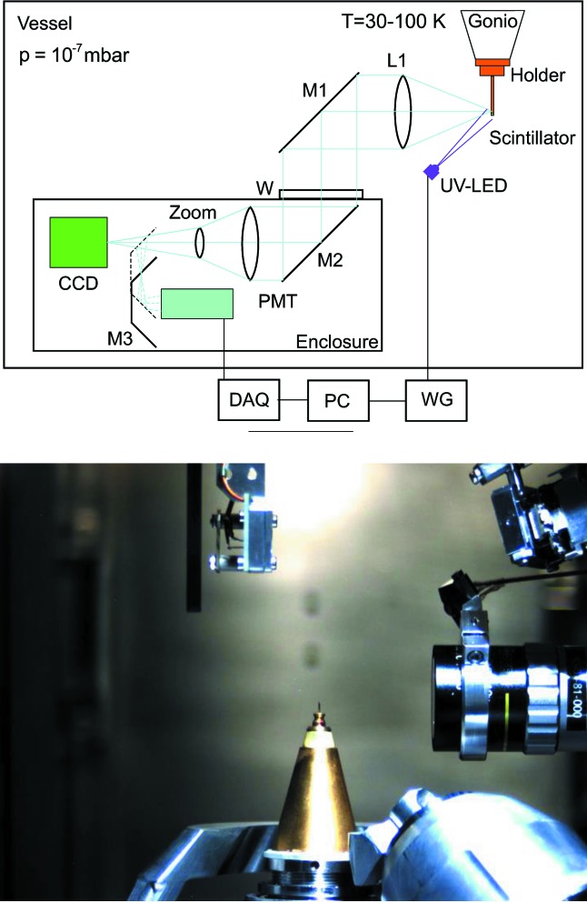
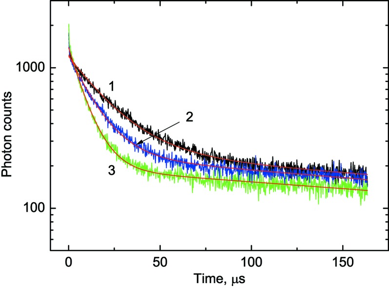
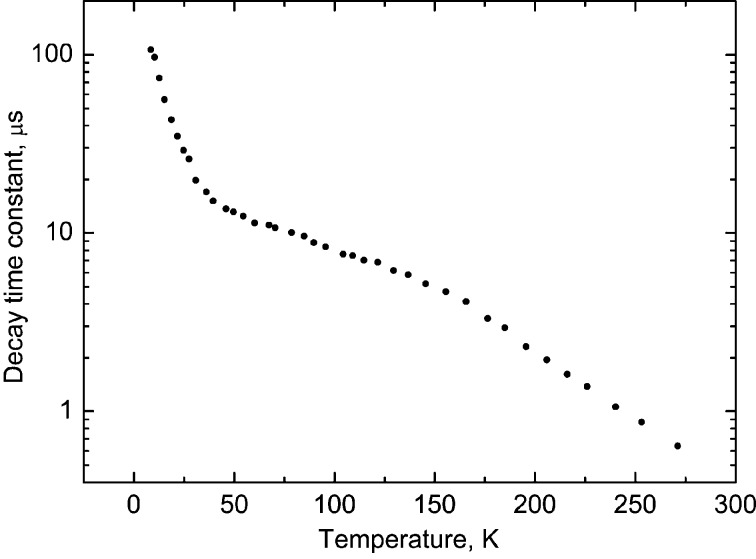
Temperature is a very important parameter when aiming to minimize radiation damage to biological samples during experiments that utilize intense ionizing radiation. A novel technique for remote, non-contact, in situ monitoring of the protein crystal temperature has been developed for the new I23 beamline at the Diamond Light Source, a facility dedicated to macromolecular crystallography (MX) with long-wavelength X-rays. The temperature is derived from the temperature-dependent decay time constant of luminescence from a minuscule scintillation sensor (<0.05 mm) located in very close proximity to the sample under test. In this work the underlying principle of cryogenic luminescence lifetime thermometry is presented, the features of the detection method and the choice of temperature sensor are discussed, and it is demonstrated how the temperature monitoring system was integrated within the viewing system of the endstation used for the visualization of protein crystals. The thermometry system was characterized using a BiGeO crystal scintillator that exhibits good responsivity of the decay time constant as a function of temperature over a wide range (8-270 K). The scintillation sensor was calibrated and the uncertainty of the temperature measurements over the primary operation temperature range of the beamline (30-150 K) was assessed to be ±1.6 K. It has been shown that the temperature of the sample holder, measured using the luminescence sensor, agrees well with the expected value. The technique was applied to characterize the thermal performance of different sample mounts that have been used in MX experiments at the I23 beamline. The thickness of the mount is shown to have the greatest impact upon the temperature distribution across the sample mount. Altogether, these tests and findings demonstrate the usefulness of the thermometry system in highlighting the challenges that remain to be addressed for the in-vacuum MX experiment to become a reliable and indispensable tool for structural biology.
在利用强电离辐射的实验中,若要将对生物样品的辐射损伤降至最低,温度是一个非常重要的参数。针对钻石光源的新I23光束线,已开发出一种用于远程、非接触、原位监测蛋白质晶体温度的新技术。该光束线是一个致力于利用长波长X射线进行大分子晶体学(MX)研究的设施。温度是通过位于被测样品非常近的位置(<0.05 mm)的一个微小闪烁传感器的发光随温度变化的衰减时间常数得出的。本文介绍了低温发光寿命测温法的基本原理,讨论了检测方法的特点和温度传感器的选择,并展示了温度监测系统是如何集成到用于蛋白质晶体可视化的终端站的观察系统中的。使用BiGeO晶体闪烁体对测温系统进行了表征,该闪烁体在很宽的温度范围(8 - 270 K)内,其衰减时间常数随温度表现出良好的响应性。对闪烁传感器进行了校准,并评估了在光束线的主要工作温度范围(30 - 150 K)内温度测量的不确定度为±1.6 K。结果表明,使用发光传感器测量的样品架温度与预期值吻合良好。该技术被应用于表征I23光束线MX实验中使用的不同样品支架的热性能。结果表明,支架的厚度对样品支架上的温度分布影响最大。总之,这些测试和结果证明了测温系统在突出真空MX实验成为结构生物学可靠且不可或缺的工具仍需解决的挑战方面的有用性。