• 文献检索
  • 文档翻译
  • 深度研究
  • 学术资讯
  • Suppr Zotero 插件Zotero 插件
  • 邀请有礼
  • 套餐&价格
  • 历史记录
应用&插件
Suppr Zotero 插件Zotero 插件浏览器插件Mac 客户端Windows 客户端微信小程序
定价
高级版会员购买积分包购买API积分包
服务
文献检索文档翻译深度研究API 文档MCP 服务
关于我们
关于 Suppr公司介绍联系我们用户协议隐私条款
关注我们

Suppr 超能文献

核心技术专利:CN118964589B侵权必究
粤ICP备2023148730 号-1Suppr @ 2026

文献检索

告别复杂PubMed语法,用中文像聊天一样搜索,搜遍4000万医学文献。AI智能推荐,让科研检索更轻松。

立即免费搜索

文件翻译

保留排版,准确专业,支持PDF/Word/PPT等文件格式,支持 12+语言互译。

免费翻译文档

深度研究

AI帮你快速写综述,25分钟生成高质量综述,智能提取关键信息,辅助科研写作。

立即免费体验

关于扩散加权乳腺成像中的分数阶微积分模型,以区分在X线筛查乳腺摄影中检测到的恶性和良性乳腺病变。

On a fractional order calculus model in diffusion weighted breast imaging to differentiate between malignant and benign breast lesions detected on X-ray screening mammography.

作者信息

Bickelhaupt Sebastian, Steudle Franziska, Paech Daniel, Mlynarska Anna, Kuder Tristan Anselm, Lederer Wolfgang, Daniel Heidi, Freitag Martin, Delorme Stefan, Schlemmer Heinz-Peter, Laun Frederik Bernd

机构信息

German Cancer Research Center (dkfz), Department of Radiology, Heidelberg, Im Neuenheimer Feld 280, Heidelberg, Germany.

German Cancer Research Center (dkfz), Medical Physics in Radiology, Heidelberg, Im Neuenheimer Feld 280, Heidelberg, Germany.

出版信息

PLoS One. 2017 Apr 28;12(4):e0176077. doi: 10.1371/journal.pone.0176077. eCollection 2017.

DOI:10.1371/journal.pone.0176077
PMID:28453516
原文链接:https://pmc.ncbi.nlm.nih.gov/articles/PMC5409173/
Abstract

OBJECTIVE

To evaluate a fractional order calculus (FROC) model in diffusion weighted imaging to differentiate between malignant and benign breast lesions in breast cancer screening work-up using recently introduced parameters (βFROC, DFROC and μFROC).

MATERIALS AND METHODS

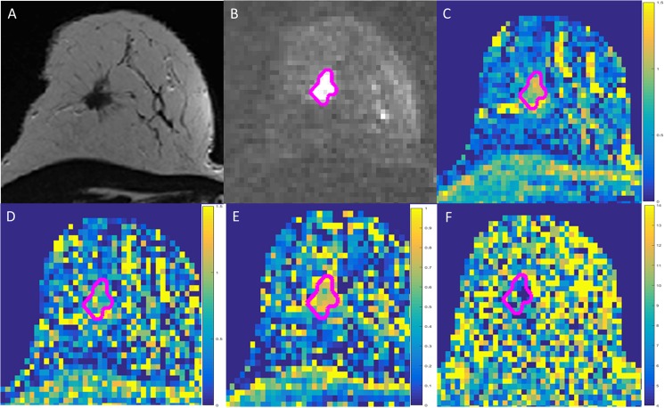
This retrospective analysis within a prospective IRB-approved study included 51 participants (mean 58.4 years) after written informed consent. All patients had suspicious screening mammograms and indication for biopsy. Prior to biopsy, full diagnostic contrast-enhanced MRI examination was acquired including diffusion-weighted-imaging (DWI, b = 0,100,750,1500 s/mm2). Conventional apparent diffusion coefficient Dapp and FROC parameters (βFROC, DFROC and μFROC) as suggested further indicators of diffusivity components were measured in benign and malignant lesions. Receiver operating characteristics (ROC) were calculated to evaluate the diagnostic performance of the parameters.

RESULTS

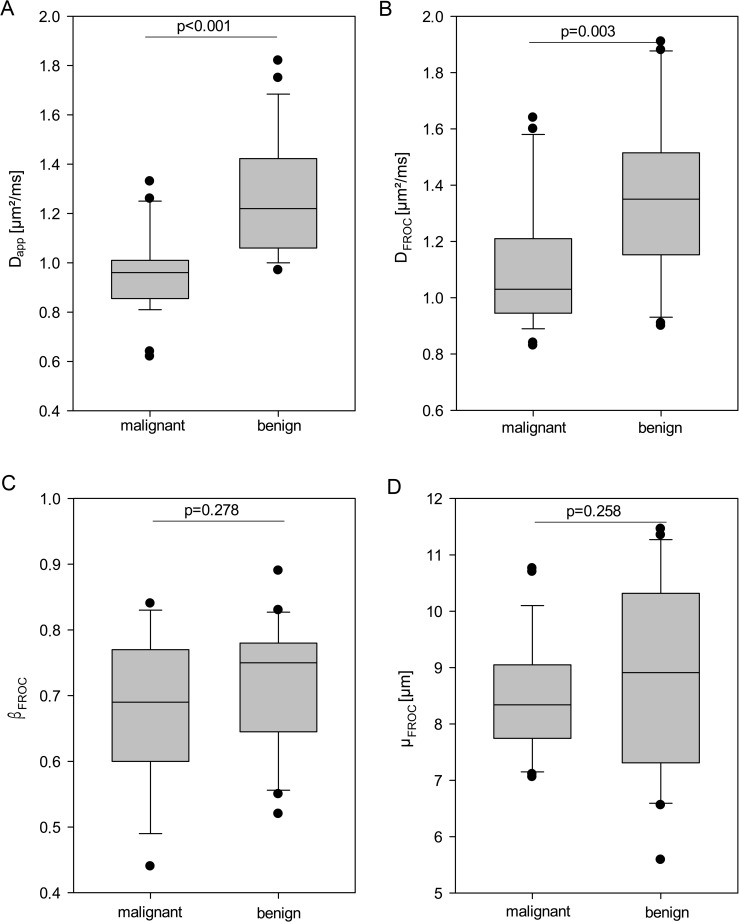
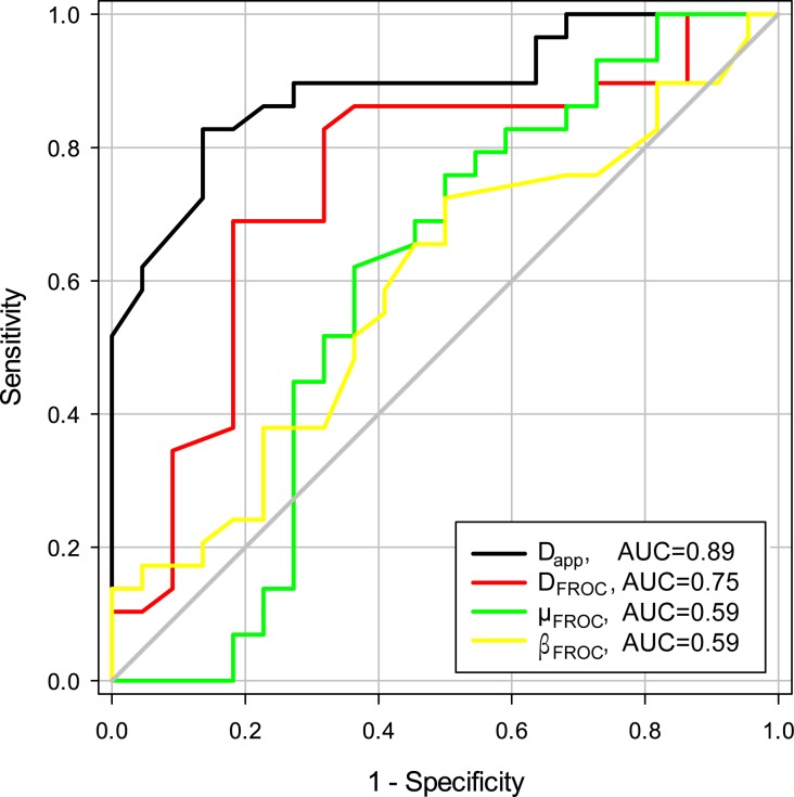
29/51 patients histopathologically revealed malignant lesions. The analysis revealed an AUC for Dapp of 0.89 (95% CI 0.80-0.98). For FROC derived parameters, AUC was 0.75 (0.60-0.89) for DFROC, 0.59 (0.43-0.75) for βFROC and 0.59 (0.42-0.77) for μFROC. Comparison of the AUC curves revealed a significantly higher AUC of Dapp compared to the FROC parameters DFROC (p = 0.009), βFROC (p = 0.003) and μFROC (p = 0.001).

CONCLUSION

In contrast to recent description in brain tumors, the apparent diffusion coefficient Dapp showed a significantly higher AUC than the recently proposed FROC parameters βFROC, DFROC and μFROC for differentiating between malignant and benign breast lesions. This might be related to the intrinsic high heterogeneity within breast tissue or to the lower maximal b-value used in our study.

摘要

目的

在扩散加权成像中评估分数阶微积分(FROC)模型,以利用最近引入的参数(βFROC、DFROC和μFROC)在乳腺癌筛查工作中区分乳腺良恶性病变。

材料与方法

这项在一项经IRB批准的前瞻性研究中的回顾性分析纳入了51名参与者(平均年龄58.4岁),所有参与者均签署了书面知情同意书。所有患者的乳腺钼靶筛查结果可疑且有活检指征。在活检前,进行了包括扩散加权成像(DWI,b = 0、100、750、1500 s/mm2)在内的全诊断性对比增强MRI检查。在良性和恶性病变中测量了传统的表观扩散系数Dapp以及作为扩散率成分进一步指标的FROC参数(βFROC、DFROC和μFROC)。计算受试者工作特征(ROC)曲线以评估这些参数的诊断性能。

结果

51例患者中29例经组织病理学检查显示为恶性病变。分析显示Dapp的曲线下面积(AUC)为0.89(95%置信区间0.80 - 0.98)。对于FROC衍生参数,DFROC的AUC为0.75(0.60 - 0.89),βFROC为0.59(0.43 - 0.75),μFROC为0.59(0.42 - 0.77)。AUC曲线比较显示,Dapp的AUC显著高于FROC参数DFROC(p = 0.009)、βFROC(p = 0.003)和μFROC(p = 0.001)。

结论

与最近关于脑肿瘤的描述不同,在区分乳腺良恶性病变方面,表观扩散系数Dapp的AUC显著高于最近提出 的FROC参数βFROC、DFROC和μFROC。这可能与乳腺组织内固有的高度异质性或本研究中使用的较低最大b值有关。

https://cdn.ncbi.nlm.nih.gov/pmc/blobs/1c0e/5409173/54fb26b55b0c/pone.0176077.g003.jpg
https://cdn.ncbi.nlm.nih.gov/pmc/blobs/1c0e/5409173/524444998b54/pone.0176077.g001.jpg
https://cdn.ncbi.nlm.nih.gov/pmc/blobs/1c0e/5409173/b8e1e6ed6432/pone.0176077.g002.jpg
https://cdn.ncbi.nlm.nih.gov/pmc/blobs/1c0e/5409173/54fb26b55b0c/pone.0176077.g003.jpg
https://cdn.ncbi.nlm.nih.gov/pmc/blobs/1c0e/5409173/524444998b54/pone.0176077.g001.jpg
https://cdn.ncbi.nlm.nih.gov/pmc/blobs/1c0e/5409173/b8e1e6ed6432/pone.0176077.g002.jpg
https://cdn.ncbi.nlm.nih.gov/pmc/blobs/1c0e/5409173/54fb26b55b0c/pone.0176077.g003.jpg

相似文献

1
On a fractional order calculus model in diffusion weighted breast imaging to differentiate between malignant and benign breast lesions detected on X-ray screening mammography.关于扩散加权乳腺成像中的分数阶微积分模型,以区分在X线筛查乳腺摄影中检测到的恶性和良性乳腺病变。
PLoS One. 2017 Apr 28;12(4):e0176077. doi: 10.1371/journal.pone.0176077. eCollection 2017.
2
Discrimination between human epidermal growth factor receptor 2 (HER2)-low-expressing and HER2-overexpressing breast cancers: a comparative study of four MRI diffusion models.人表皮生长因子受体 2(HER2)低表达与过表达乳腺癌的鉴别:四种 MRI 扩散模型的比较研究。
Eur Radiol. 2024 Apr;34(4):2546-2559. doi: 10.1007/s00330-023-10198-x. Epub 2023 Sep 6.
3
Comparison of continuous-time random walk and fractional order calculus models in characterizing breast lesions using histogram analysis.连续时间随机游走模型与分数阶微积分模型在利用直方图分析表征乳腺病变中的比较
Magn Reson Imaging. 2024 May;108:47-58. doi: 10.1016/j.mri.2024.01.012. Epub 2024 Feb 1.
4
Value of fractional-order calculus (FROC) model diffusion-weighted imaging combined with simultaneous multi-slice (SMS) acceleration technology for evaluating benign and malignant breast lesions.分数阶微积分(FROC)模型扩散加权成像联合同时多切片(SMS)加速技术评估乳腺良恶性病变的价值。
BMC Med Imaging. 2024 Jul 29;24(1):190. doi: 10.1186/s12880-024-01368-4.
5
Differentiation of benign and malignant breast lesions using diffusion-weighted imaging with a fractional-order calculus model.应用分数阶微积分模型的扩散加权成像对良恶性乳腺病变的鉴别诊断。
Eur J Radiol. 2023 Feb;159:110646. doi: 10.1016/j.ejrad.2022.110646. Epub 2022 Dec 15.
6
Prediction of malignancy by a radiomic signature from contrast agent-free diffusion MRI in suspicious breast lesions found on screening mammography.基于对比剂-free 扩散 MRI 的放射组学特征预测筛查性乳腺钼靶摄影中可疑乳腺病变的恶性程度。
J Magn Reson Imaging. 2017 Aug;46(2):604-616. doi: 10.1002/jmri.25606. Epub 2017 Feb 2.
7
Intravoxel incoherent motion (IVIM) in evaluation of breast lesions: comparison with conventional DWI.体素内不相干运动(IVIM)在评估乳腺病变中的应用:与常规 DWI 的比较。
Eur J Radiol. 2013 Dec;82(12):e782-9. doi: 10.1016/j.ejrad.2013.08.006. Epub 2013 Aug 13.
8
Monoexponential, Biexponential, and stretched-exponential models using diffusion-weighted imaging: A quantitative differentiation of breast lesions at 3.0T.基于扩散加权成像的单指数、双指数和拉伸指数模型:3.0T 下乳腺病变的定量鉴别。
J Magn Reson Imaging. 2019 Nov;50(5):1461-1467. doi: 10.1002/jmri.26729. Epub 2019 Mar 27.
9
Diffusion tensor imaging for characterizing tumor microstructure and improving diagnostic performance on breast MRI: a prospective observational study.弥散张量成像用于表征肿瘤微结构并提高乳腺 MRI 的诊断性能:一项前瞻性观察研究。
Breast Cancer Res. 2019 Sep 4;21(1):102. doi: 10.1186/s13058-019-1183-3.
10
Differentiation of salivary gland tumor using diffusion-weighted imaging with a fractional order calculus model.应用分数阶微积分模型的扩散加权成像对唾液腺肿瘤进行鉴别诊断。
Br J Radiol. 2020 Sep 1;93(1113):20200052. doi: 10.1259/bjr.20200052. Epub 2020 Jul 10.

引用本文的文献

1
Respiratory abnormalities in sarcoidosis: physiopathology and early diagnosis using oscillometry combined with respiratory modeling.结节病中的呼吸异常:结合振荡法与呼吸模型的病理生理学及早期诊断
BMC Pulm Med. 2025 Feb 7;25(1):68. doi: 10.1186/s12890-025-03510-6.
2
Swarm learning with weak supervision enables automatic breast cancer detection in magnetic resonance imaging.基于弱监督的群体学习实现磁共振成像中乳腺癌的自动检测。
Commun Med (Lond). 2025 Feb 6;5(1):38. doi: 10.1038/s43856-024-00722-5.
3
Multimodal apparent diffusion MRI model in noninvasive evaluation of breast cancer and Ki-67 expression.

本文引用的文献

1
Prediction of malignancy by a radiomic signature from contrast agent-free diffusion MRI in suspicious breast lesions found on screening mammography.基于对比剂-free 扩散 MRI 的放射组学特征预测筛查性乳腺钼靶摄影中可疑乳腺病变的恶性程度。
J Magn Reson Imaging. 2017 Aug;46(2):604-616. doi: 10.1002/jmri.25606. Epub 2017 Feb 2.
2
Differentiation of Low- and High-Grade Gliomas Using High b-Value Diffusion Imaging with a Non-Gaussian Diffusion Model.使用非高斯扩散模型的高b值扩散成像鉴别低级别和高级别胶质瘤
AJNR Am J Neuroradiol. 2016 Sep;37(9):1643-9. doi: 10.3174/ajnr.A4836. Epub 2016 Jun 2.
3
Independent value of image fusion in unenhanced breast MRI using diffusion-weighted and morphological T2-weighted images for lesion characterization in patients with recently detected BI-RADS 4/5 x-ray mammography findings.
多模态表观扩散 MRI 模型在无创评估乳腺癌及 Ki-67 表达中的应用。
Cancer Imaging. 2024 Oct 11;24(1):137. doi: 10.1186/s40644-024-00780-x.
4
Value of fractional-order calculus (FROC) model diffusion-weighted imaging combined with simultaneous multi-slice (SMS) acceleration technology for evaluating benign and malignant breast lesions.分数阶微积分(FROC)模型扩散加权成像联合同时多切片(SMS)加速技术评估乳腺良恶性病变的价值。
BMC Med Imaging. 2024 Jul 29;24(1):190. doi: 10.1186/s12880-024-01368-4.
5
Stability of Radiomic Features against Variations in Lesion Segmentations Computed on Apparent Diffusion Coefficient Maps of Breast Lesions.基于乳腺病变表观扩散系数图计算的病变分割变化下放射组学特征的稳定性
Diagnostics (Basel). 2024 Jul 3;14(13):1427. doi: 10.3390/diagnostics14131427.
6
Diffusion-Weighted Imaging for Skin Pathologies of the Breast-A Feasibility Study.乳腺皮肤病变的扩散加权成像——一项可行性研究
Diagnostics (Basel). 2024 Apr 29;14(9):934. doi: 10.3390/diagnostics14090934.
7
Radiomics to Differentiate Malignant and Benign Breast Lesions: A Systematic Review and Diagnostic Test Accuracy Meta-Analysis.基于影像组学鉴别乳腺良恶性病变:一项系统评价与诊断试验准确性的Meta分析
Cureus. 2023 Nov 18;15(11):e49015. doi: 10.7759/cureus.49015. eCollection 2023 Nov.
8
Discrimination between human epidermal growth factor receptor 2 (HER2)-low-expressing and HER2-overexpressing breast cancers: a comparative study of four MRI diffusion models.人表皮生长因子受体 2(HER2)低表达与过表达乳腺癌的鉴别:四种 MRI 扩散模型的比较研究。
Eur Radiol. 2024 Apr;34(4):2546-2559. doi: 10.1007/s00330-023-10198-x. Epub 2023 Sep 6.
9
Cervical Carcinoma: Evaluation Using Diffusion MRI With a Fractional Order Calculus Model and its Correlation With Histopathologic Findings.宫颈癌:使用分数阶微积分模型的扩散磁共振成像评估及其与组织病理学结果的相关性
Front Oncol. 2022 Apr 5;12:851677. doi: 10.3389/fonc.2022.851677. eCollection 2022.
10
Oscillation Mechanics, Integer and Fractional Respiratory Modeling in COPD: Effect of Obstruction Severity.COPD 中的振荡力学、整数和分数呼吸建模:阻塞严重程度的影响。
Int J Chron Obstruct Pulmon Dis. 2020 Dec 8;15:3273-3289. doi: 10.2147/COPD.S276690. eCollection 2020.
在未增强乳腺MRI中,利用扩散加权成像和形态学T2加权成像进行图像融合,对近期乳腺影像报告和数据系统(BI-RADS)4/5级乳腺钼靶X线摄影检查结果的患者进行病变特征分析的独立价值。
Eur Radiol. 2017 Feb;27(2):562-569. doi: 10.1007/s00330-016-4400-9. Epub 2016 May 18.
4
Quantification of microcirculatory parameters by joint analysis of flow-compensated and non-flow-compensated intravoxel incoherent motion (IVIM) data.通过对流动补偿和非流动补偿体素内不相干运动(IVIM)数据进行联合分析来量化微循环参数。
NMR Biomed. 2016 May;29(5):640-9. doi: 10.1002/nbm.3505. Epub 2016 Mar 8.
5
[Selected clinically established and scientific techniques of diffusion-weighted MRI. In the context of imaging in oncology].[扩散加权磁共振成像的选定临床成熟及科学技术。在肿瘤学成像背景下]
Radiologe. 2016 Feb;56(2):137-47. doi: 10.1007/s00117-015-0066-6.
6
Apparent diffusion coefficient in differentiation between malignant and benign breast masses: does size matter?表观扩散系数在乳腺良恶性肿块鉴别中的应用:大小有影响吗?
Clin Radiol. 2016 Feb;71(2):170-7. doi: 10.1016/j.crad.2015.11.006. Epub 2015 Dec 11.
7
Fast and Noninvasive Characterization of Suspicious Lesions Detected at Breast Cancer X-Ray Screening: Capability of Diffusion-weighted MR Imaging with MIPs.快速无创特征分析乳腺癌 X 射线筛查中可疑病变:扩散加权磁共振成像 MIPs 的能力。
Radiology. 2016 Mar;278(3):689-97. doi: 10.1148/radiol.2015150425. Epub 2015 Sep 29.
8
Unenhanced breast MRI (STIR, T2-weighted TSE, DWIBS): An accurate and alternative strategy for detecting and differentiating breast lesions.乳腺磁共振成像平扫(短T1反转恢复序列、快速自旋回波T2加权成像、背景抑制扩散加权成像):一种用于检测和鉴别乳腺病变的准确且可替代的策略。
Magn Reson Imaging. 2015 Oct;33(8):951-5. doi: 10.1016/j.mri.2015.06.002. Epub 2015 Jun 25.
9
Breast-cancer screening--viewpoint of the IARC Working Group.乳腺癌筛查——国际癌症研究机构工作组的观点
N Engl J Med. 2015 Jun 11;372(24):2353-8. doi: 10.1056/NEJMsr1504363. Epub 2015 Jun 3.
10
Differentiation of Low- and High-Grade Pediatric Brain Tumors with High b-Value Diffusion-weighted MR Imaging and a Fractional Order Calculus Model.利用高b值扩散加权磁共振成像和分数阶微积分模型鉴别儿童低级别和高级别脑肿瘤
Radiology. 2015 Nov;277(2):489-96. doi: 10.1148/radiol.2015142156. Epub 2015 Jun 2.