Zhang Ting, Hu Yuan, Wang Ting, Cai Peiling
Department of Medical Cell Biology and Genetics, Southwest Medical University, Luzhou, Sichuan 646000, P.R. China.
Department of Anatomy and Histology, School of Medicine, Chengdu University, Chengdu, Sichuan 610106, P.R. China.
Int J Mol Med. 2017 Jul;40(1):21-30. doi: 10.3892/ijmm.2017.2980. Epub 2017 May 9.
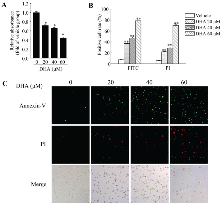
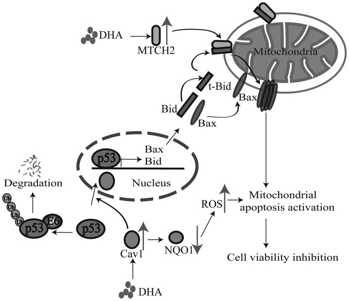
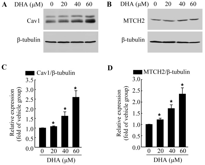
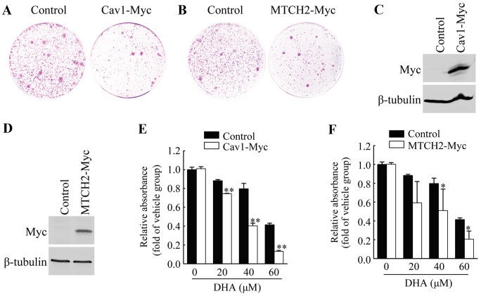
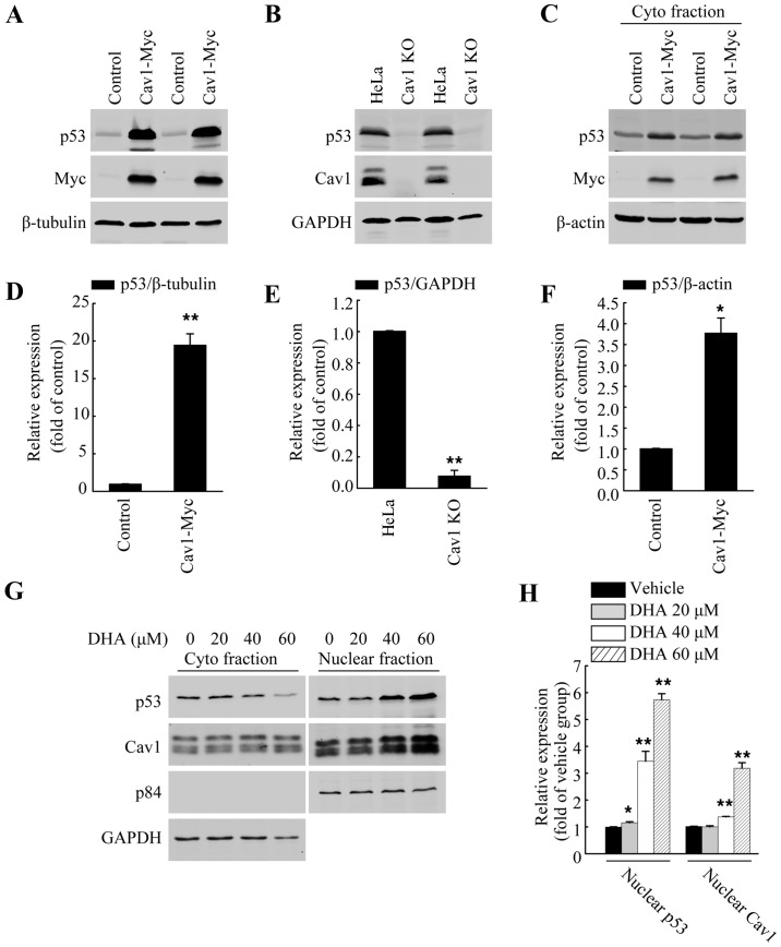
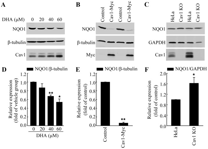
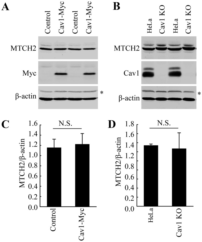
Dihydroartemisinin (DHA) has been shown to inhibit the viability of various cancer cells. Previous studies have revealed that the mechanisms involved in the inhibitory effects of DHA are based on theactivation of p53 and the mitochondrial-related cell death pathway. However, the exact association between upstream signaling and the activation of cell death pathway remains unclear. In this study, we found that DHA treatment induced the upregulation of caveolin 1 (Cav1) and mitochondrial carrier homolog 2 (MTCH2) in HeLa cells, and this was associated with the DHA-induced inhibition of cell viability and DHA-induced apoptosis. Additionally, the overexpression of Cav1 and MTCH2 in HeLa cells enhanced the inhibitory effects of DHA on cell viability. Moreover, we also found that the upregulation of Cav1 contributed to the DHA-mediated p53 activation and the downregulation of the redox enzyme, NAD(P)H:quinone oxidoreductase 1 (NQO1), which have been reported to contribute to the activation of the cell death pathway. Of note, we also found that DHA induced the nuclear translocation and accumulation of both Cav1 and p53, indicating a novel potential mechanism, namely the regulation of p53 activation by Cav1. On the whole, our study identified Cav1 and MTCH2 as the molecular targets of DHA and revealed a new link between the upstream Cav1/MTCH2 upregulation and the downstream activation of the cell death pathway involved in the DHA-mediated inhibition of cell viability.
双氢青蒿素(DHA)已被证明可抑制多种癌细胞的活力。先前的研究表明,DHA抑制作用的机制基于p53的激活和线粒体相关的细胞死亡途径。然而,上游信号与细胞死亡途径激活之间的确切关联仍不清楚。在本研究中,我们发现DHA处理可诱导HeLa细胞中小窝蛋白1(Cav1)和线粒体载体同源物2(MTCH2)的上调,这与DHA诱导的细胞活力抑制和DHA诱导的细胞凋亡相关。此外,HeLa细胞中Cav1和MTCH2的过表达增强了DHA对细胞活力的抑制作用。此外,我们还发现Cav1的上调有助于DHA介导的p53激活以及氧化还原酶NAD(P)H:醌氧化还原酶1(NQO1)的下调,据报道这有助于细胞死亡途径的激活。值得注意的是,我们还发现DHA诱导了Cav1和p53的核转位和积累,表明一种新的潜在机制,即Cav1对p53激活的调节。总体而言,我们的研究确定Cav1和MTCH2为DHA的分子靶点,并揭示了上游Cav1/MTCH2上调与DHA介导的细胞活力抑制所涉及的细胞死亡途径下游激活之间的新联系。