• 文献检索
  • 文档翻译
  • 深度研究
  • 学术资讯
  • Suppr Zotero 插件Zotero 插件
  • 邀请有礼
  • 套餐&价格
  • 历史记录
应用&插件
Suppr Zotero 插件Zotero 插件浏览器插件Mac 客户端Windows 客户端微信小程序
定价
高级版会员购买积分包购买API积分包
服务
文献检索文档翻译深度研究API 文档MCP 服务
关于我们
关于 Suppr公司介绍联系我们用户协议隐私条款
关注我们

Suppr 超能文献

核心技术专利:CN118964589B侵权必究
粤ICP备2023148730 号-1Suppr @ 2026

文献检索

告别复杂PubMed语法,用中文像聊天一样搜索,搜遍4000万医学文献。AI智能推荐,让科研检索更轻松。

立即免费搜索

文件翻译

保留排版,准确专业,支持PDF/Word/PPT等文件格式,支持 12+语言互译。

免费翻译文档

深度研究

AI帮你快速写综述,25分钟生成高质量综述,智能提取关键信息,辅助科研写作。

立即免费体验

颈动脉分叉处的高帧率矢量血流成像。

High-frame rate vector flow imaging of the carotid bifurcation.

作者信息

Goddi Alfredo, Bortolotto Chandra, Fiorina Ilaria, Raciti Maria Vittoria, Fanizza Marianna, Turpini Elena, Boffelli Giulia, Calliada Fabrizio

机构信息

Centro Medico SME-Diagnostica per Immagini, 31, Via L. Pirandello, 21100, Varese, VA, Italy.

Radiology Department, Fondazione IRCCS Policlinico San Matteo Pavia, Pavia, Italy.

出版信息

Insights Imaging. 2017 Jun;8(3):319-328. doi: 10.1007/s13244-017-0554-5. Epub 2017 May 12.

DOI:10.1007/s13244-017-0554-5
PMID:28500487
原文链接:https://pmc.ncbi.nlm.nih.gov/articles/PMC5438320/
Abstract

UNLABELLED

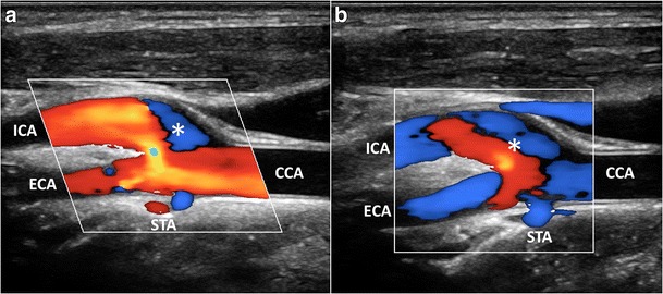
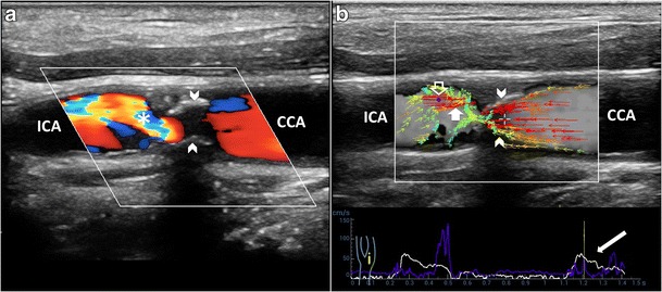
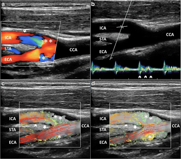
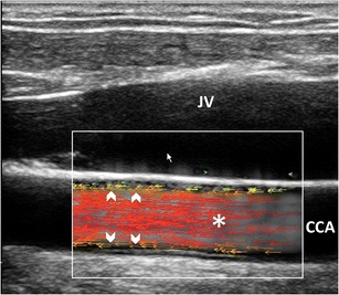
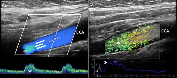
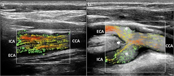
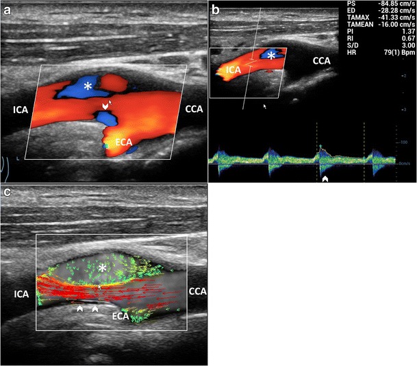
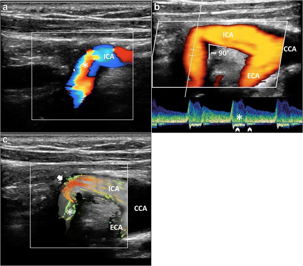
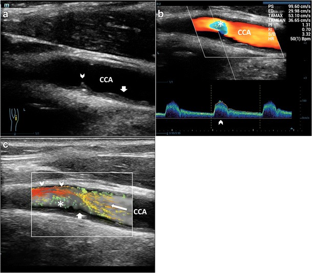
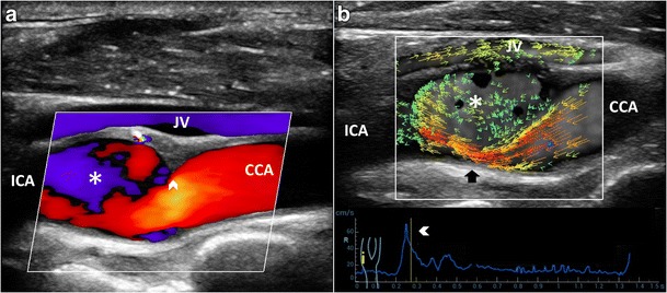
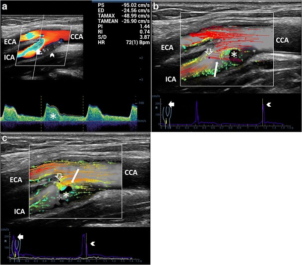
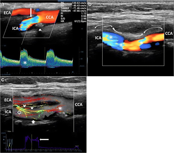
Carotid artery atherosclerotic disease is still a significant cause of cerebrovascular morbidity and mortality. A new angle-independent technique, measuring and visualizing blood flow velocities in all directions, called vector flow imaging (VFI) is becoming available from several vendors. VFI can provide more intuitive and quantitative imaging of vortex formation, which is not clearly distinguishable in the color Doppler image. VFI, as quantitative method assessing disturbed flow patterns of the carotid bifurcation, has the potential to allow better understanding of the diagnostic value of complex flow and to enhance risk stratification. This pictorial review article will show which new information VFI adds for the knowledge of hemodynamics in comparison to the conventional ultrasound techniques.

TEACHING POINTS

• VFI is an angle-independent technique measuring flow velocities in all directions. • This kind of VFI is based on a plane wave multidirectional excitation technique. • VFI allows quantitative assessment of carotid streamlines progression and visualizes vorticity. • VFI does not allow a precise comprehension of streamlines' 3D shape. • VFI allows a better understanding of carotid artery complex flows.

摘要

未标注

颈动脉粥样硬化疾病仍是脑血管发病和死亡的重要原因。一种新的与角度无关的技术,即测量和可视化各个方向血流速度的矢量血流成像(VFI),正由多家供应商提供。VFI能够提供更直观、定量的涡旋形成成像,这在彩色多普勒图像中无法清晰分辨。作为评估颈动脉分叉处紊乱血流模式的定量方法,VFI有可能更好地理解复杂血流的诊断价值并加强风险分层。这篇图片综述文章将展示与传统超声技术相比,VFI为血流动力学知识增添了哪些新信息。

教学要点

• VFI是一种测量各个方向血流速度的与角度无关的技术。• 这种VFI基于平面波多向激发技术。• VFI允许对颈动脉流线进展进行定量评估并可视化涡度。• VFI无法精确理解流线的三维形状。• VFI有助于更好地理解颈动脉复杂血流。

https://cdn.ncbi.nlm.nih.gov/pmc/blobs/f5da/5438320/8d8bb58603cf/13244_2017_554_Fig12_HTML.jpg
https://cdn.ncbi.nlm.nih.gov/pmc/blobs/f5da/5438320/dfe78a52361d/13244_2017_554_Fig1_HTML.jpg
https://cdn.ncbi.nlm.nih.gov/pmc/blobs/f5da/5438320/39e68eb8d01b/13244_2017_554_Fig2_HTML.jpg
https://cdn.ncbi.nlm.nih.gov/pmc/blobs/f5da/5438320/8b1e35e70fd4/13244_2017_554_Fig3_HTML.jpg
https://cdn.ncbi.nlm.nih.gov/pmc/blobs/f5da/5438320/f5ed60f41804/13244_2017_554_Fig4_HTML.jpg
https://cdn.ncbi.nlm.nih.gov/pmc/blobs/f5da/5438320/55afd7a2fa59/13244_2017_554_Fig5_HTML.jpg
https://cdn.ncbi.nlm.nih.gov/pmc/blobs/f5da/5438320/058293bcabb4/13244_2017_554_Fig6_HTML.jpg
https://cdn.ncbi.nlm.nih.gov/pmc/blobs/f5da/5438320/e9661c5bdf07/13244_2017_554_Fig7_HTML.jpg
https://cdn.ncbi.nlm.nih.gov/pmc/blobs/f5da/5438320/0dd81fb43e0d/13244_2017_554_Fig8_HTML.jpg
https://cdn.ncbi.nlm.nih.gov/pmc/blobs/f5da/5438320/15745093dca3/13244_2017_554_Fig9_HTML.jpg
https://cdn.ncbi.nlm.nih.gov/pmc/blobs/f5da/5438320/35b1b302baf9/13244_2017_554_Fig10_HTML.jpg
https://cdn.ncbi.nlm.nih.gov/pmc/blobs/f5da/5438320/7456e7f4792c/13244_2017_554_Fig11_HTML.jpg
https://cdn.ncbi.nlm.nih.gov/pmc/blobs/f5da/5438320/8d8bb58603cf/13244_2017_554_Fig12_HTML.jpg

相似文献

1
High-frame rate vector flow imaging of the carotid bifurcation.颈动脉分叉处的高帧率矢量血流成像。
Insights Imaging. 2017 Jun;8(3):319-328. doi: 10.1007/s13244-017-0554-5. Epub 2017 May 12.
2
Precise evaluation of blood flow patterns in human carotid bifurcation based on high-frame-rate vector flow imaging.基于高帧率血流向量成像的人体颈动脉分叉处血流模式的精确评估。
J Clin Ultrasound. 2023 Jul-Aug;51(6):1070-1077. doi: 10.1002/jcu.23489. Epub 2023 May 18.
3
High-Frame Rate Vector Flow Imaging of the Carotid Bifurcation in Healthy Adults: Comparison With Color Doppler Imaging.健康成年人颈动脉分叉处的高帧率矢量血流成像:与彩色多普勒成像的比较
J Ultrasound Med. 2018 Sep;37(9):2263-2275. doi: 10.1002/jum.14579. Epub 2018 Mar 25.
4
Accuracy and Precision of a Plane Wave Vector Flow Imaging Method in the Healthy Carotid Artery.平面波矢量血流成像方法在健康颈动脉中的准确性和精确性
Ultrasound Med Biol. 2018 Aug;44(8):1727-1741. doi: 10.1016/j.ultrasmedbio.2018.03.017. Epub 2018 May 4.
5
Velocity vector comparison between vector flow imaging and computational fluid dynamics in the carotid bifurcation.颈动脉分叉处血流向量成像与计算流体动力学的速度向量比较。
Ultrasonics. 2023 Feb;128:106860. doi: 10.1016/j.ultras.2022.106860. Epub 2022 Oct 8.
6
Ultrasound vector projectile imaging for detection of altered carotid bifurcation hemodynamics during reductions in cardiac output.超声向量抛射成像用于检测心输出量降低期间颈动脉分叉处血流动力学的改变。
Med Phys. 2020 Feb;47(2):431-440. doi: 10.1002/mp.13905. Epub 2019 Dec 4.
7
Vector flow imaging techniques: An innovative ultrasonographic technique for the study of blood flow.矢量血流成像技术:一种用于研究血流的创新性超声技术。
J Clin Ultrasound. 2017 Nov 12;45(9):582-588. doi: 10.1002/jcu.22519. Epub 2017 Jul 21.
8
First experiences of ultrasound vector flow imaging at the femoropopliteal artery in peripheral arterial disease.股腘动脉周围动脉疾病中超声向量血流成像的初步经验。
Vasa. 2023 Nov;52(6):394-401. doi: 10.1024/0301-1526/a001095. Epub 2023 Oct 17.
9
A Comparison Study of Vector Velocity, Spectral Doppler and Magnetic Resonance of Blood Flow in the Common Carotid Artery.颈总动脉血流的矢量速度、频谱多普勒及磁共振对比研究
Ultrasound Med Biol. 2018 Aug;44(8):1751-1761. doi: 10.1016/j.ultrasmedbio.2018.05.002. Epub 2018 May 24.
10
Efficacy of ultrasound vector flow imaging in tracking omnidirectional pulsatile flow.超声矢量血流成像在追踪全方向搏动血流中的效能
Med Phys. 2023 Mar;50(3):1699-1714. doi: 10.1002/mp.16168. Epub 2023 Jan 14.

引用本文的文献

1
Management and Treatment of Carotid Stenosis: Overview of Therapeutic Possibilities and Comparison Between Interventional Radiology, Surgery and Hybrid Procedure.颈动脉狭窄的管理与治疗:治疗选择概述及介入放射学、外科手术与杂交手术的比较
Diagnostics (Basel). 2025 Jul 1;15(13):1679. doi: 10.3390/diagnostics15131679.
2
Plaque biomarkers from high-frame rate vector flow imaging and shear wave elastography in mild carotid stenosis.高帧率矢量血流成像和剪切波弹性成像在轻度颈动脉狭窄中的斑块生物标志物
Ultrasonography. 2025 Jul;44(4):274-284. doi: 10.14366/usg.25031. Epub 2025 May 6.
3
Possibility of modern ultrasound imaging of portal venous system.

本文引用的文献

1
Novel flow quantification of the carotid bulb and the common carotid artery with vector flow ultrasound.利用矢量血流超声对颈动脉球部和颈总动脉进行新型血流定量分析。
Ultrasound Med Biol. 2014 Nov;40(11):2700-6. doi: 10.1016/j.ultrasmedbio.2014.06.001. Epub 2014 Sep 11.
2
Vector projectile imaging: time-resolved dynamic visualization of complex flow patterns.矢量射弹成像:复杂流动模式的时间分辨动态可视化
Ultrasound Med Biol. 2014 Sep;40(9):2295-309. doi: 10.1016/j.ultrasmedbio.2014.03.014. Epub 2014 Jun 25.
3
Combined vector velocity and spectral Doppler imaging for improved imaging of complex blood flow in the carotid arteries.
门静脉系统现代超声成像的可能性。
Clin Hemorheol Microcirc. 2025 May;90(1):3-13. doi: 10.1177/13860291251324086. Epub 2025 Jun 2.
4
Imaging of Carotid Stenosis: Where Are We Standing? Comparison of Multiparametric Ultrasound, CT Angiography, and MRI Angiography, with Recent Developments.颈动脉狭窄的影像学检查:我们目前的状况如何?多参数超声、CT血管造影和MRI血管造影的比较及最新进展
Diagnostics (Basel). 2024 Aug 6;14(16):1708. doi: 10.3390/diagnostics14161708.
5
Ultrasound Speckle Decorrelation Analysis-Based Velocimetry for 3D-Velocity-Components Measurement Using a 1D Transducer Array.基于超声散斑去相关分析的一维线阵换能器三维速度分量测量速度法。
Adv Sci (Weinh). 2024 Aug;11(32):e2401173. doi: 10.1002/advs.202401173. Epub 2024 Jun 20.
6
Quantitative evaluation using carotid ultrasonography-based high-frame-rate vector flow imaging in patients with low carotid stenosis.基于颈动脉超声的高帧率矢量血流成像对轻度颈动脉狭窄患者的定量评估
Br J Radiol. 2024 Aug 1;97(1160):1476-1482. doi: 10.1093/bjr/tqae115.
7
Evaluafion of the efficacy of wall shear stress in carotid artery stenting.颈动脉支架置入术中壁面剪应力疗效评估。 不过你原文中“Evaluafion”拼写有误,正确的是“Evaluation” 。
Heliyon. 2024 May 18;10(11):e31383. doi: 10.1016/j.heliyon.2024.e31383. eCollection 2024 Jun 15.
8
Associations between carotid plaque shape, biomechanical parameters, and ischemic stroke in mild carotid stenosis with a single plaque.轻度颈动脉狭窄伴单发斑块时颈动脉斑块形状、生物力学参数与缺血性卒中之间的关联
Ultrasonography. 2024 May;43(3):209-219. doi: 10.14366/usg.24019. Epub 2024 Apr 2.
9
Clinical Application of High-Frame-Rate Vector Flow Imaging in Evaluation of Carotid Atherosclerotic Stenosis.高帧率矢量血流成像在评估颈动脉粥样硬化狭窄中的临床应用
Diagnostics (Basel). 2023 Jan 31;13(3):519. doi: 10.3390/diagnostics13030519.
10
Quantitative Evaluation of Post-stenotic Blood Flow Disturbance in Canine Femoral Artery Stenosis Model: An Early Experience With Vector Flow Imaging.犬股动脉狭窄模型中狭窄后血流紊乱的定量评估:矢量血流成像的早期经验
Front Cardiovasc Med. 2022 Feb 24;9:829825. doi: 10.3389/fcvm.2022.829825. eCollection 2022.
联合矢量速度和频谱多普勒成像以改善颈动脉复杂血流成像。
Ultrasound Med Biol. 2014 Jul;40(7):1629-40. doi: 10.1016/j.ultrasmedbio.2014.01.021. Epub 2014 Apr 27.
4
Real-time vector velocity assessment through multigate Doppler and plane waves.实时向量速度评估通过多门控多普勒和平面波。
IEEE Trans Ultrason Ferroelectr Freq Control. 2014 Feb;61(2):314-24. doi: 10.1109/TUFFC.2014.6722616.
5
Ultrasonic colour Doppler imaging.超声彩色多谱勒成像。
Interface Focus. 2011 Aug 6;1(4):490-502. doi: 10.1098/rsfs.2011.0017. Epub 2011 May 6.
6
Emerging trends in CV flow visualization.CV 流可视化的新兴趋势。
JACC Cardiovasc Imaging. 2012 Mar;5(3):305-16. doi: 10.1016/j.jcmg.2012.01.003.
7
Grading carotid stenosis using ultrasonic methods.使用超声方法对颈动脉狭窄进行分级。
Stroke. 2012 Mar;43(3):916-21. doi: 10.1161/STROKEAHA.111.636084. Epub 2012 Feb 16.
8
Comparison of real-time in vivo spectral and vector velocity estimation.实时体内光谱和向量速度估计的比较。
Ultrasound Med Biol. 2012 Jan;38(1):145-51. doi: 10.1016/j.ultrasmedbio.2011.10.003. Epub 2011 Nov 21.
9
Comprehensive 4D velocity mapping of the heart and great vessels by cardiovascular magnetic resonance.心血管磁共振全面 4D 速度图绘制心脏和大血管。
J Cardiovasc Magn Reson. 2011 Jan 14;13(1):7. doi: 10.1186/1532-429X-13-7.
10
Role of hemodynamic shear stress in cardiovascular disease.血流切应力在心血管疾病中的作用。
Atherosclerosis. 2011 Feb;214(2):249-56. doi: 10.1016/j.atherosclerosis.2010.09.008. Epub 2010 Sep 18.