Bagheri Mohammad H, Ahlman Mark A, Lindenberg Liza, Turkbey Baris, Lin Jeffrey, Cahid Civelek Ali, Malayeri Ashkan A, Agarwal Piyush K, Choyke Peter L, Folio Les R, Apolo Andrea B
Clinical Image Processing Service, Radiology and Imaging Sciences Department, Clinical Center, National Institutes of Health, Bethesda, MD.
Nuclear Medicine Department, Clinical Center, National Institutes of Health, Bethesda, MD; Radiology and Imaging Sciences Department, Clinical Center, National Institutes of Health, Bethesda, MD.
Urol Oncol. 2017 Jul;35(7):473-491. doi: 10.1016/j.urolonc.2017.04.014. Epub 2017 May 12.
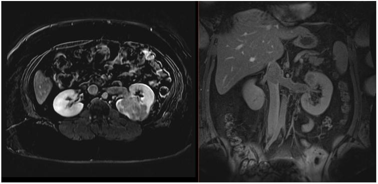
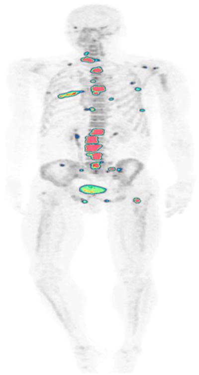
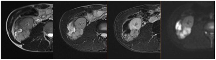
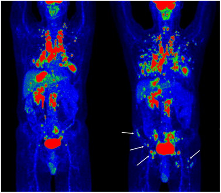
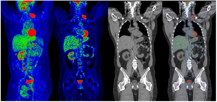
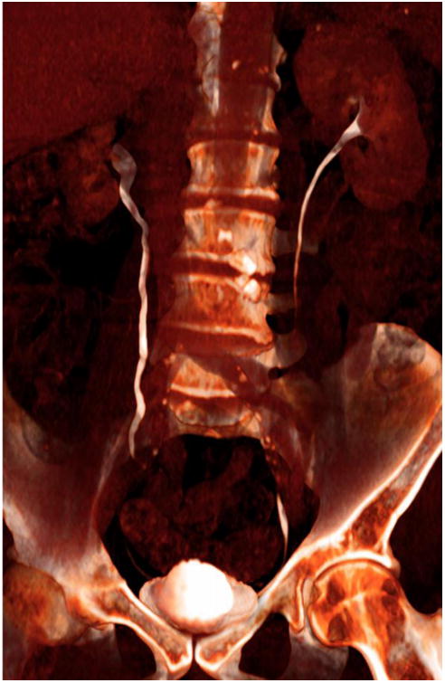
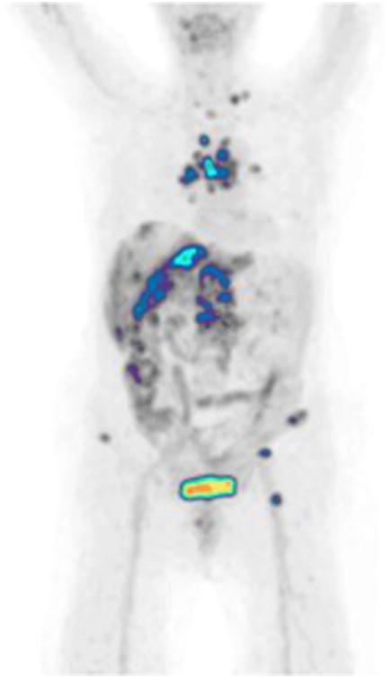
Medical imaging of the 3 most common genitourinary (GU) cancers-prostate adenocarcinoma, renal cell carcinoma, and urothelial carcinoma of the bladder-has evolved significantly during the last decades. The most commonly used imaging modalities for the diagnosis, staging, and follow-up of GU cancers are computed tomography, magnetic resonance imaging (MRI), and positron emission tomography (PET). Multiplanar multidetector computed tomography and multiparametric MRI with diffusion-weighted imaging are the main imaging modalities for renal cell carcinoma and urothelial carcinoma, and although multiparametric MRI is rapidly becoming the main imaging tool in the evaluation of prostate adenocarcinoma, biopsy is still required for diagnosis. Functional and molecular imaging using 18-fluorodeoxyglucose-PET and sodium fluoride-PET are essential for the diagnosis, and especially follow-up, of metastatic GU tumors. This review provides an overview of the latest advances in the imaging of these 3 major GU cancers.
在过去几十年中,三种最常见的泌尿生殖系统(GU)癌症——前列腺腺癌、肾细胞癌和膀胱尿路上皮癌的医学成像技术有了显著发展。用于GU癌症诊断、分期和随访的最常用成像方式是计算机断层扫描(CT)、磁共振成像(MRI)和正电子发射断层扫描(PET)。多平面多探测器计算机断层扫描和具有扩散加权成像的多参数MRI是肾细胞癌和膀胱尿路上皮癌的主要成像方式,尽管多参数MRI正迅速成为评估前列腺腺癌的主要成像工具,但诊断仍需活检。使用18-氟脱氧葡萄糖-PET和氟化钠-PET的功能和分子成像对于转移性GU肿瘤的诊断,尤其是随访至关重要。本综述概述了这三种主要GU癌症成像的最新进展。