Cornelis Francois H, Durack Jeremy C, Kimm Simon Y, Wimmer Thomas, Coleman Jonathan A, Solomon Stephen B, Srimathveeravalli Govindarajan
Department of Radiology, Memorial Sloan-Kettering Cancer Center, 1275 York Avenue, New York, NY, 10065, USA.
Department of Urology, Palo Alto Medical Foundation, Palo Alto, CA, USA.
Cardiovasc Intervent Radiol. 2017 Oct;40(10):1600-1608. doi: 10.1007/s00270-017-1692-3. Epub 2017 May 17.
To compare ablation boundary sharpness after percutaneous radiofrequency ablation (RFA), cryoablation (CA), microwave ablation (MWA) and irreversible electroporation (IRE) ablation in normal swine liver and kidney.
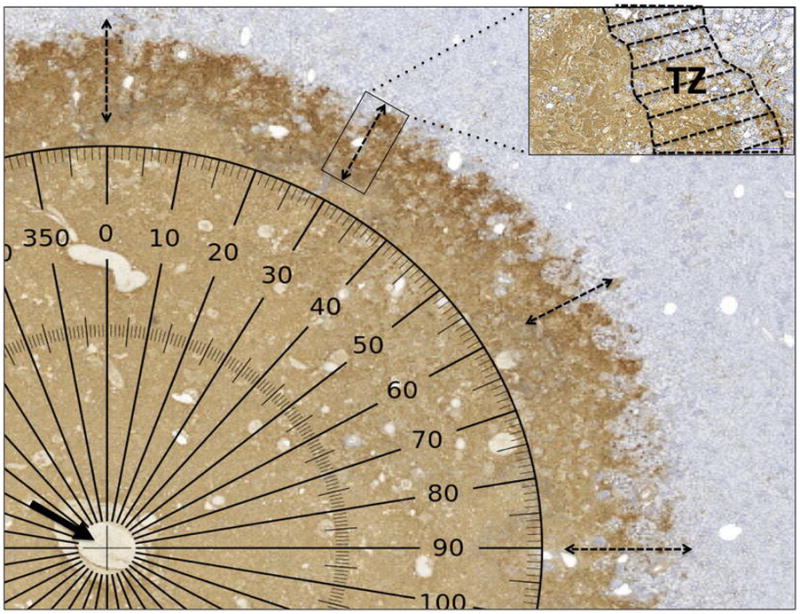
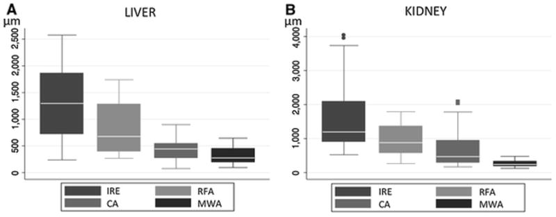
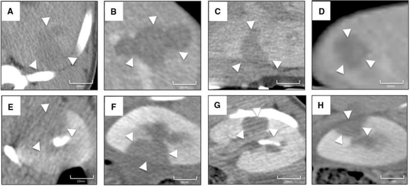
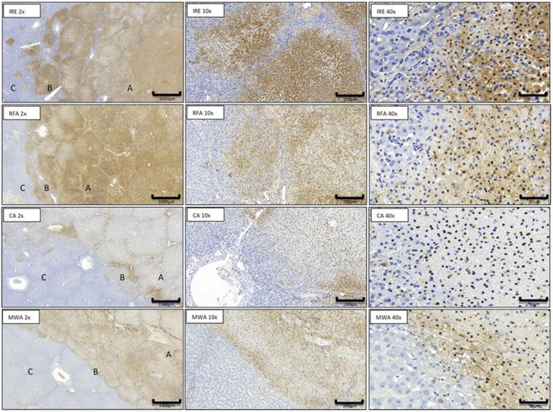
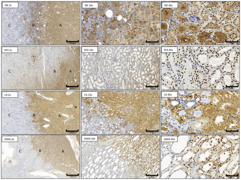
Percutaneous CT-guided RFA (n = 5), CA (n = 5), MWA (n = 5) and IRE (n = 5) were performed in the liver and kidney of four Yorkshire pigs. Parameters were chosen to produce ablations 2-3 cm in diameter with a single ablation probe. Contrast-enhanced CT imaging was performed 24 h after ablation, and animals were killed. Treated organs were removed and processed for histologic analysis with hematoxylin and eosin, and terminal deoxynucleotidyl transferase dUTP nick end labeling (TUNEL). Three readers independently analyzed CT, H&E and TUNEL stained images of the ablation boundary to delineate regions of (1) viable cells, (2) complete necrosis or (3) mixture of viable and necrotic cells which was defined as the transition zone (TZ). The width of TZ was compared across the techniques and organs.
Ablations appeared as non-contrast-enhancing regions on CT with sharp transition to enhancing normal tissue. On TUNEL stained slides, the mean width (μm) of the TZ after MWA was 319 ± 157 in liver and 267 ± 95 in kidney, which was significantly lower than RFA (811 ± 477 and 938 ± 429); CA (452 ± 222 and 700 ± 563); and IRE (1319 ± 682 and 1570 ± 962) (all p < 0.01). No significant differences were observed between the organs.
Under similar conditions, the width of the TZ at the ablation boundary varies significantly between different ablation techniques.
比较经皮射频消融(RFA)、冷冻消融(CA)、微波消融(MWA)和不可逆电穿孔(IRE)消融在正常猪肝和肾中的消融边界清晰度。
对4只约克夏猪的肝脏和肾脏进行经皮CT引导下的RFA(n = 5)、CA(n = 5)、MWA(n = 5)和IRE(n = 5)。选择参数以使用单个消融探针产生直径2 - 3厘米的消融灶。消融后24小时进行对比增强CT成像,然后处死动物。取出处理过的器官,用苏木精和伊红以及末端脱氧核苷酸转移酶dUTP缺口末端标记法(TUNEL)进行组织学分析。三位阅片者独立分析消融边界的CT、苏木精和伊红染色以及TUNEL染色图像,以描绘出(1)活细胞、(2)完全坏死或(3)活细胞与坏死细胞混合区域(定义为过渡区(TZ))。比较不同技术和器官的TZ宽度。
消融灶在CT上表现为无对比增强区域,与增强的正常组织有清晰的过渡。在TUNEL染色切片上,MWA后肝脏TZ的平均宽度(μm)为319 ± 157,肾脏为267 ± 95,显著低于RFA(811 ± 477和938 ± 429);CA(452 ± 222和700 ± 563);以及IRE(1319 ± 682和1570 ± 962)(所有p < 0.01)。各器官之间未观察到显著差异。
在相似条件下,不同消融技术的消融边界TZ宽度差异显著。