Berninger Markus T, Mohajerani Pouyan, Wildgruber Moritz, Beziere Nicolas, Kimm Melanie A, Ma Xiaopeng, Haller Bernhard, Fleming Megan J, Vogt Stephan, Anton Martina, Imhoff Andreas B, Ntziachristos Vasilis, Meier Reinhard, Henning Tobias D
Department of Orthopaedic Sports Medicine, Klinikum rechts der Isar, Technische Universität München, Munich, Germany.
Department of Trauma and Orthopaedic Surgery, BG Unfallklinik Murnau, Murnau, Germany.
Photoacoustics. 2017 May 4;6:37-47. doi: 10.1016/j.pacs.2017.04.002. eCollection 2017 Jun.
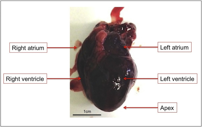
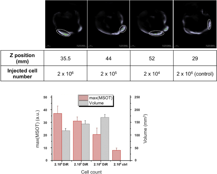
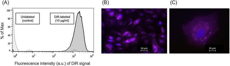
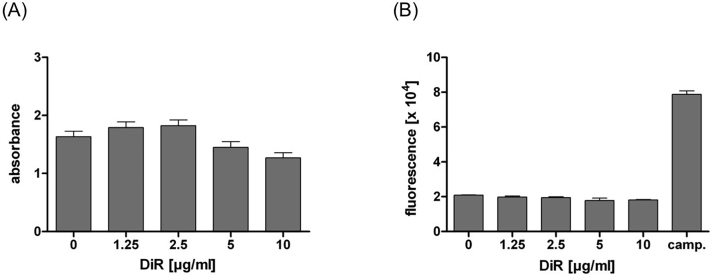
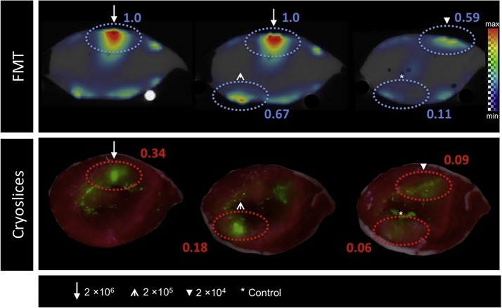
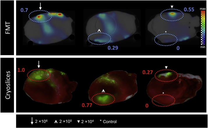
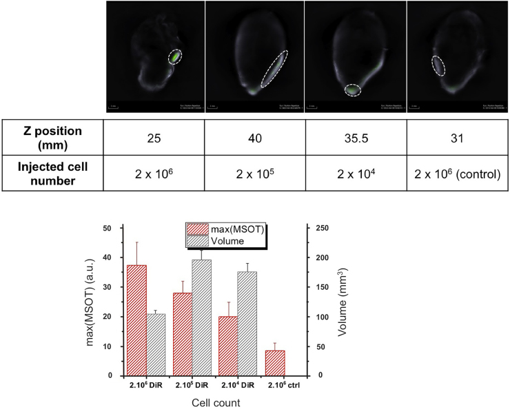
The distribution of intramyocardially injected rabbit MSCs, labeled with the near-infrared dye 1,1'-dioctadecyl-3,3,3',3'-tetramethylindotricarbo-cyanine-iodide (DiR) using hybrid Fluorescence Molecular Tomography-X-ray Computed Tomography (FMT-XCT) and Multispectral Optoacoustic Tomography (MSOT) imaging technologies, was investigated. Viability and induction of apoptosis of DiR labeled MSCs were assessed by XTT- and Caspase-3/-7-testing . 2 × 10, 2 × 10 and 2 × 10 MSCs labeled with 5 and 10 μg DiR/ml were injected into fresh frozen rabbit hearts. FMT-XCT, MSOT and fluorescence cryosection imaging were performed. Concentrations up to 10 μg DiR/ml did not cause apoptosis (p > 0.05). FMT and MSOT imaging of labeled MSCs led to a strong signal. The imaging modalities highlighted a difference in cell distribution and concentration correlated to the number of injected cells. cryosectioning confirmed the molecular fluorescence signal. FMT and MSOT are sensitive imaging techniques offering high-anatomic resolution in terms of detection and distribution of intramyocardially injected stem cells in a rabbit model.
利用混合荧光分子断层扫描-X射线计算机断层扫描(FMT-XCT)和多光谱光声断层扫描(MSOT)成像技术,研究了用近红外染料1,1'-二辛基-3,3,3',3'-四甲基吲哚三碳菁碘化物(DiR)标记的兔心肌内注射间充质干细胞(MSCs)的分布情况。通过XTT和Caspase-3/-7检测评估DiR标记的MSCs的活力和凋亡诱导情况。将用5和10μg DiR/ml标记的2×10⁶、2×10⁶和2×10⁶个MSCs注射到新鲜冷冻的兔心脏中。进行了FMT-XCT、MSOT和荧光冷冻切片成像。高达10μg DiR/ml的浓度不会导致细胞凋亡(p>0.05)。标记的MSCs的FMT和MSOT成像产生了强烈信号。这些成像方式突出了与注射细胞数量相关的细胞分布和浓度差异。冷冻切片证实了分子荧光信号。FMT和MSOT是敏感的成像技术,在兔模型中对心肌内注射干细胞的检测和分布提供了高解剖分辨率。