Lemaster Jeanne E, Chen Fang, Kim Taeho, Hariri Ali, Jokerst Jesse V
Department of NanoEngineering, San Diego (UCSD), 9500 Gilman Drive, La Jolla, California 92093, United States.
Materials Science and Engineering Program, San Diego (UCSD), 9500 Gilman Drive, La Jolla, California 92093, United States.
ACS Appl Nano Mater. 2018 Mar 23;1(3):1321-1331. doi: 10.1021/acsanm.8b00063. Epub 2018 Mar 2.
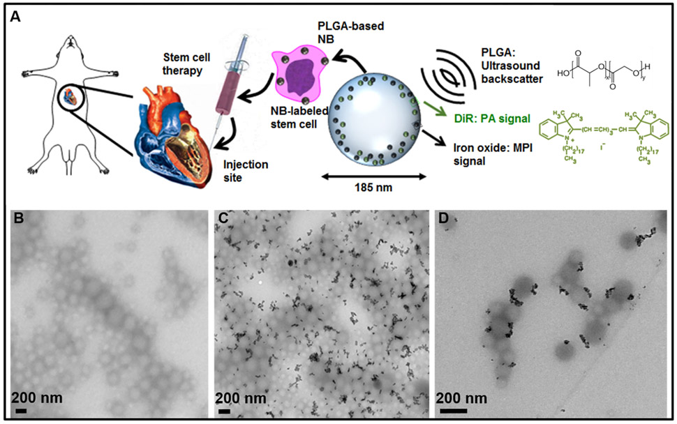
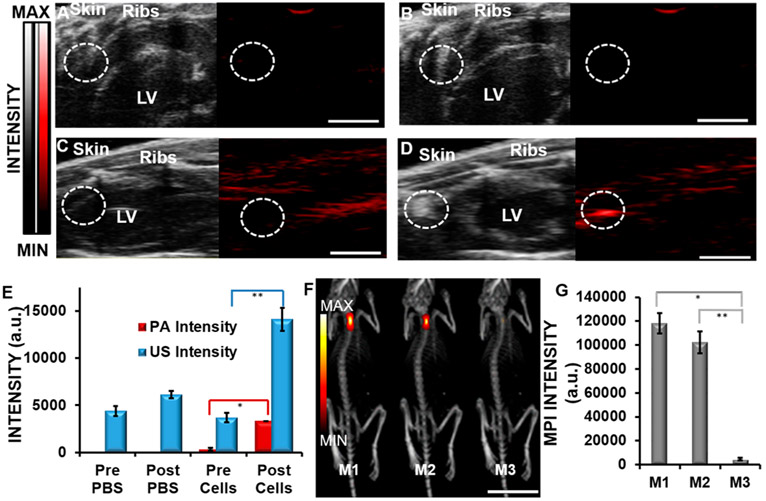
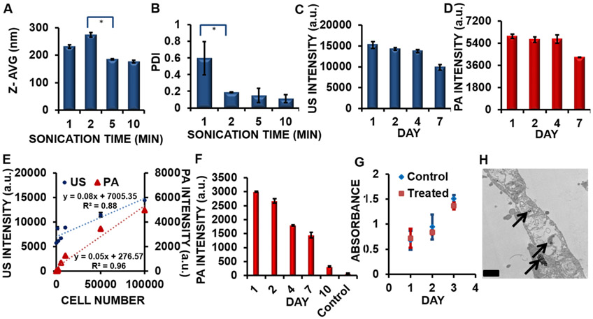
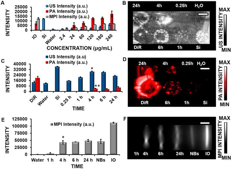
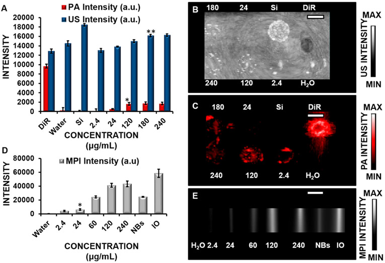
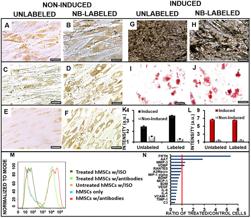
Stem cell therapy has the potential to improve tissue remodeling and repair. For cardiac stem cell therapy, methods to improve the injection and tracking of stem cells may help to increase patient outcomes. Here we describe a multimodal approach that combines ultrasound imaging, photoacoustic imaging, and magnetic particle imaging (MPI). Ultrasound imaging offers real-time guidance, photoacoustic imaging offers enhanced contrast, and MPI offers high-contrast, deep-tissue imaging. This work was facilitated by a poly(lactic--glycolic acid) (PLGA)-based iron oxide nanobubble labeled with 1,1'-dioctadecyl-3,3,3',3'-tetramethylindotricarbocyanine iodide (DiR) as a trimodal contrast agent. The PLGA coating facilitated the ultrasound signal, the DiR increased the photoacoustic signal, and the iron oxide facilitated the MPI signal. We confirmed that cell metabolism, proliferation, differentiation, and migration were not adversely affected by cell treatment with nanobubbles. The nanobubble-labeled cells were injected intramyocardially into live mice for real-time imaging. Ultrasound imaging showed a 3.8-fold increase in the imaging intensity of labeled cells postinjection compared to the baseline; photoacoustic imaging showed a 10.2-fold increase in the cardiac tissue signal postinjection. The MPI intensity of the nanobubble-treated human mesenchymal stem cells injected into the hearts of mice was approximately 20-fold greater than the negative control.
干细胞疗法有改善组织重塑和修复的潜力。对于心脏干细胞疗法而言,改进干细胞注射和追踪的方法可能有助于提高患者的治疗效果。在此,我们描述一种结合超声成像、光声成像和磁粒子成像(MPI)的多模态方法。超声成像提供实时引导,光声成像提供增强的对比度,而MPI提供高对比度的深部组织成像。这项工作借助一种基于聚乳酸-乙醇酸共聚物(PLGA)的氧化铁纳米气泡得以推进,该纳米气泡用1,1'-二辛基-3,3,3',3'-四甲基吲哚三碳菁碘化物(DiR)标记,作为一种三模态造影剂。PLGA涂层增强了超声信号,DiR增强了光声信号,而氧化铁增强了MPI信号。我们证实,纳米气泡处理细胞对细胞代谢、增殖、分化和迁移没有不利影响。将纳米气泡标记的细胞心肌内注射到活小鼠体内进行实时成像。超声成像显示,注射后标记细胞的成像强度相比基线增加了3.8倍;光声成像显示,注射后心脏组织信号增加了10.2倍。注射到小鼠心脏中的纳米气泡处理的人间充质干细胞的MPI强度比阴性对照大约高20倍。