Ysasi Alexandra B, Wagner Willi L, Valenzuela Cristian D, Kienzle Arne, Servais Andrew B, Bennett Robert D, Tsuda Akira, Ackermann Maximilian, Mentzer Steven J
Laboratory of Adaptive and Regenerative Biology, Brigham & Women's Hospital, Harvard Medical School, Boston, Massachusetts, United States of America.
Institute of Functional and Clinical Anatomy, University Medical Center of the Johannes Gutenberg-University, Mainz, Germany.
PLoS One. 2017 May 19;12(5):e0177921. doi: 10.1371/journal.pone.0177921. eCollection 2017.
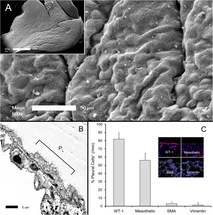
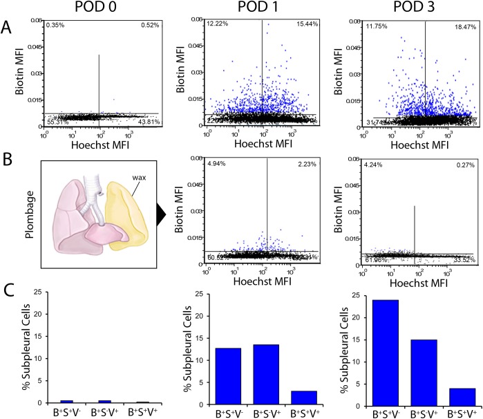
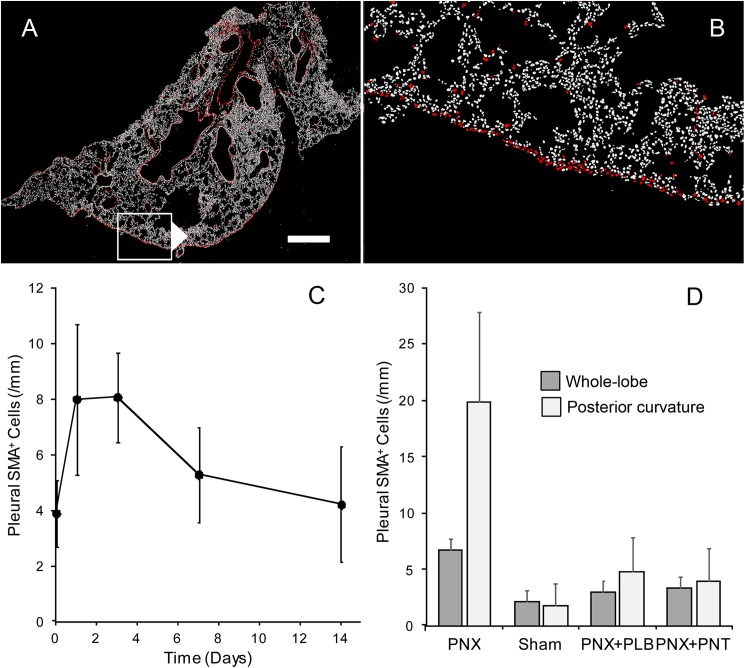
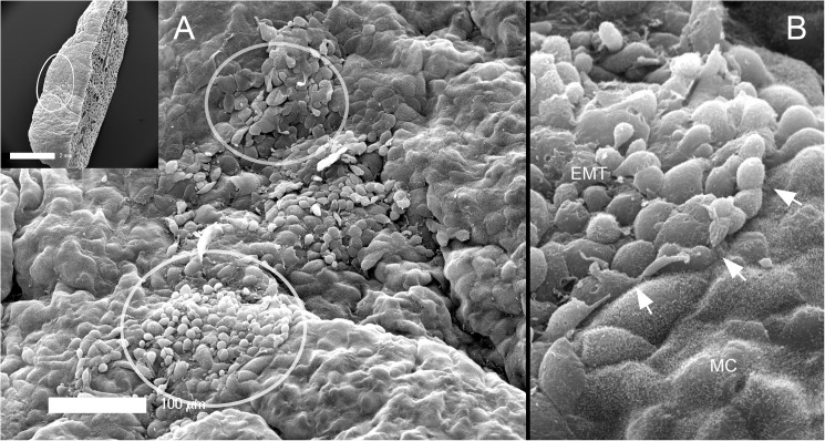
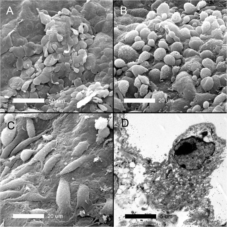
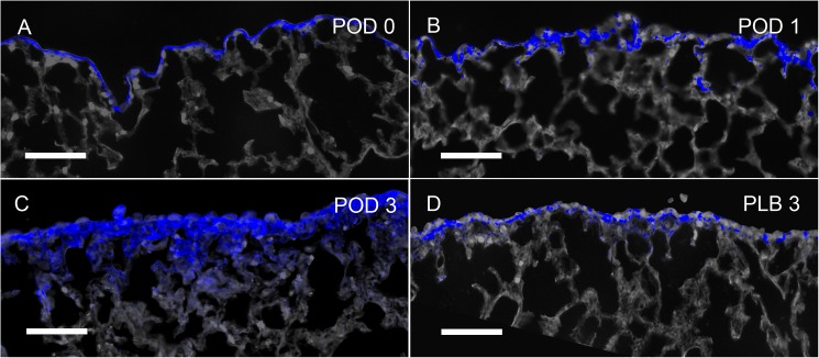
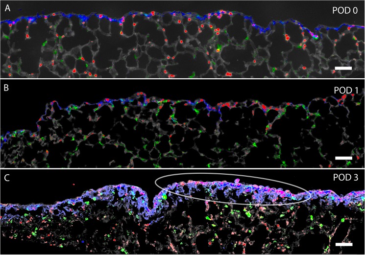
In many mammals, including rodents and humans, removal of one lung results in the compensatory growth of the remaining lung; however, the mechanism of compensatory lung growth is unknown. Here, we investigated the changes in morphology and phenotype of pleural cells after pneumonectomy. Between days 1 and 3 after pneumonectomy, cells expressing α-smooth muscle actin (SMA), a cytoplasmic marker of myofibroblasts, were significantly increased in the pleura compared to surgical controls (p < .01). Scanning electron microscopy of the pleural surface 3 days post-pneumonectomy demonstrated regions of the pleura with morphologic features consistent with epithelial-mesenchymal transition (EMT); namely, cells with disrupted intercellular junctions and an acquired mesenchymal (rounded and fusiform) morphotype. To detect the migration of the transitional pleural cells into the lung, a biotin tracer was used to label the pleural mesothelial cells at the time of surgery. By post-operative day 3, image cytometry of post-pneumonectomy subpleural alveoli demonstrated a 40-fold increase in biotin+ cells relative to pneumonectomy-plus-plombage controls (p < .01). Suggesting a similar origin in space and time, the distribution of cells expressing biotin, SMA, or vimentin demonstrated a strong spatial autocorrelation in the subpleural lung (p < .001). We conclude that post-pneumonectomy compensatory lung growth involves EMT with the migration of transitional mesothelial cells into subpleural alveoli.
在包括啮齿动物和人类在内的许多哺乳动物中,切除一侧肺会导致剩余肺的代偿性生长;然而,代偿性肺生长的机制尚不清楚。在此,我们研究了肺切除术后胸膜细胞形态和表型的变化。肺切除术后第1天至第3天,与手术对照组相比,胸膜中表达α平滑肌肌动蛋白(SMA,成肌纤维细胞的细胞质标志物)的细胞显著增加(p < 0.01)。肺切除术后3天对胸膜表面进行扫描电子显微镜检查,发现胸膜区域具有与上皮-间质转化(EMT)一致的形态学特征;即细胞间连接破坏且获得间充质(圆形和梭形)形态的细胞。为了检测过渡性胸膜细胞向肺内的迁移,在手术时使用生物素示踪剂标记胸膜间皮细胞。到术后第3天,肺切除术后胸膜下肺泡的图像细胞术显示,与肺切除加填充对照组相比,生物素阳性细胞增加了40倍(p < 0.01)。表达生物素、SMA或波形蛋白的细胞分布在胸膜下肺中显示出强烈的空间自相关性(p < 0.001),这表明在空间和时间上有相似的起源。我们得出结论,肺切除术后代偿性肺生长涉及EMT以及过渡性间皮细胞向胸膜下肺泡的迁移。