Kienzle Arne, Servais Andrew B, Ysasi Alexandra B, Gibney Barry C, Valenzuela Cristian D, Wagner Willi L, Ackermann Maximilian, Mentzer Steven J
Laboratory of Adaptive and Regenerative Biology, Brigham & Women's Hospital, Harvard Medical School, Boston, MA, United States.
Department of Diagnostic and Interventional Radiology, Translational Lung Research Center Heidelberg (TLRC), Member of German Center for Lung Research (DZL), University of Heidelberg, Heidelberg, Germany.
Front Med (Lausanne). 2018 Apr 5;5:89. doi: 10.3389/fmed.2018.00089. eCollection 2018.
The mesothelium, the surface layer of the heart, lung, bowel, liver, and tunica vaginalis, is a complex tissue implicated in organ-specific diseases and regenerative biology; however, the mechanism of mesothelial repair after surgical injury is unknown. Previous observations indicated seeding of denuded mesothelium by free-floating mesothelial cells may contribute to mesothelial healing. In this study, we investigated the prevalence of mesothelial cells in pleural fluid during the 7 days following pulmonary surgery.
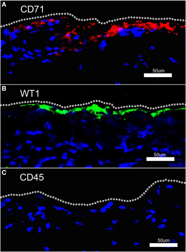
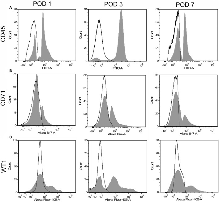
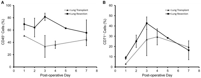
Flow cytometry was employed to study pleural fluid of 45 patients after lung resection or transplantation. We used histologically validated mesothelial markers (CD71 and WT1) to estimate the prevalence of mesothelial cells.
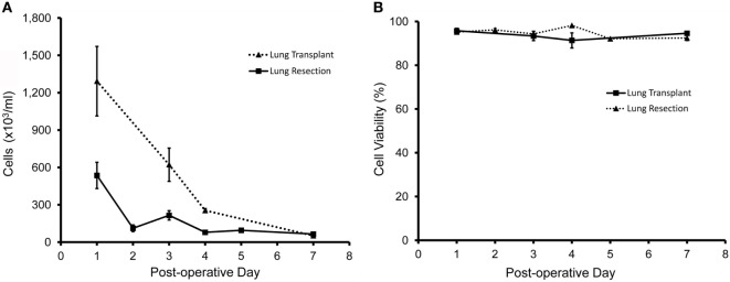
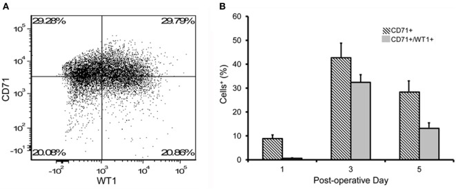
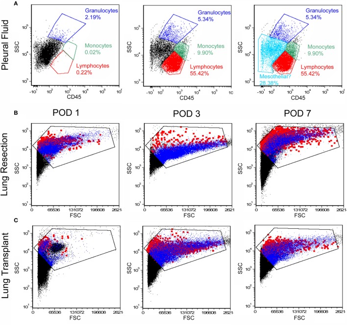
The viability of pleural fluid cells approached 100%. Leukocytes and mesothelial cells were identified in the pleural fluid within the first week after surgery. The leukocyte concentration was relatively stable at all time points. In contrast, mesothelial cells, identified by CD71 and WT1 peaked on POD3. The broad expression of CD71 molecule in postoperative pleural fluid suggests that many of the free-floating non-leukocyte cells were activated or proliferative mesothelial cells.
We demonstrated that pleural fluid post lung surgery is a source of mesothelial cells; most of these cells appear to be viable and, as shown by CD71 staining, activated mesothelial cells. The observed peak of mesothelial cells on POD3 is consistent with a potential reparative role of free-floating mesothelial cells after pulmonary surgery.
间皮是心脏、肺、肠、肝脏和鞘膜的表层,是一种与器官特异性疾病和再生生物学相关的复杂组织;然而,手术损伤后间皮修复的机制尚不清楚。先前的观察表明,游离的间皮细胞播种到剥脱的间皮上可能有助于间皮愈合。在本研究中,我们调查了肺手术后7天内胸腔积液中间皮细胞的发生率。
采用流式细胞术研究45例肺切除或移植患者的胸腔积液。我们使用经组织学验证的间皮标志物(CD71和WT1)来估计间皮细胞的发生率。
胸腔积液细胞的活力接近100%。术后第一周内胸腔积液中可识别出白细胞和间皮细胞。白细胞浓度在所有时间点相对稳定。相比之下,通过CD71和WT1识别的间皮细胞在术后第3天达到峰值。CD71分子在术后胸腔积液中的广泛表达表明,许多游离的非白细胞细胞是活化的或增殖的间皮细胞。
我们证明肺手术后的胸腔积液是间皮细胞的一个来源;这些细胞中的大多数似乎是有活力的,并且如CD71染色所示,是活化的间皮细胞。观察到的术后第3天间皮细胞峰值与肺手术后游离间皮细胞的潜在修复作用一致。